Canalblog
Suivre ce blog Administration + Créer mon blog

Maladie de GAUCHER : actualités

Archives
Derniers commentaires
15 mars 2013

Le paradigme de la maladie de Gaucher : du réticulum endoplasmitque aux comorbidités

La maladie de Gaucher est caractérisée par une accumulation de glucosylcéramides dans les lysosomes, liée à une diminution de l’activité de la béta-glucocérébrosidase acide. Cette baisse d’activité dépend de la quantité d’enzyme mutante mature qui parvient aux lysosomes et elle est modulée par l’importance de la dégradation de chaque variant par la voie ERAD du réticulum endoplasmique. L’ERAD est un processus complexe qui régule l’identification des protéines mal repliées dans le réticulum endoplasmique, leurs tentatives de repliage, leur sortie du réticulum endoplasmique vers le cytoplasme, leur poly-ubiquitination et leur dégradation dans le protéasome. Toutes ces étapes mettent en jeu un grand nombre de protéines, dont certaines sont des protéines chaperonnes calcium-dépendantes du réticulum tandis que d’autres participent à la poly-ubiquitination. Les taux de calcium et de cholestérol du réticulum endoplasmique ainsi que d’autres facteurs non encore identifiés déterminent le degré d’ERAD des différents variants mutants de la béta-glucocérébrosidase acide. Pour les auteurs, ce processus joue un rôle important dans le développement de la maladie de Parkinson, et d’autres maladies, chez les porteurs et les patients qui présentent une maladie de Gaucher. Les variants mutants de la béta-glucocérébrosidase subissent une poly-ubiquitination à laquelle participent les ligases E3: celles-ci sont alors détournées de leur activité sur d’autres substrats cellulaires, ce qui annule des processus cellulaires normaux.

OB

Le paradigme de la maladie de Gaucher : du réticulum endoplasmique aux comorbidités

Publicité
Publicité
15 mars 2013

Une approche thérapeutique innovante de la maladie de Gaucher

 
Dans la maladie de Gaucher, l’enzymothérapie substitutive a prouvé son efficacité sur les manifestations systémiques mais la taille de l’enzyme de supplémentation ne lui permet pas de traverser la barrière hémato-encéphalique et de traiter l’atteinte du système nerveux central. Pendant de nombreuses années, il a été admis que les anomalies de la séquence des acides aminés de la glucocérébrosidase diminuaient son activité catalytique. Il a été récemment démontré que la baisse de son activité résulte d’une diminution quantitative de l’expression de cette enzyme, ce qui implique l’existence d’un processus spécifique au cours duquel les polypeptides mal repliés sont ciblés par des médiateurs de l’homéostasie protéique pour être dégradés. La modulation de l’action de ces médiateurs pourrait prévenir la dégradation de la glucocérébrosidase et lui rendre sa fonction. Des études récentes ont montré que de petites molécules inhibitrices des histones déacétylases (qui peuvent traverser la barrière hémato-encéphalique) étaient capables de moduler ces médiateurs dans des cellules provenant de patients présentant une maladie de Gaucher et d’augmenter la quantité et l’activité de la glucocérébrosidase.
OB

An innovative approach to the treatment of Gaucher disease and possibly other metabolic disorders of the brain
[24-08-2012]
Roscoe O. Brady1, Chunzhang Yang2 and Zhengping Zhuang2
1 Scientist Emeritus, National Institutes of Health, Building 10 Room 3D03, Bethesda, MD 20892-1260, USA.
2 Surgical Neurology Branch, National Institutes of Health, Building 10 Room 7N246, Bethesda, MD 20892-1414, USA.

Abstract  

The extraordinary benefit of enzyme replacement therapy (ERT) on the systemic manifestations of Gaucher disease was demonstrated in 1991. Since that time, investigators have devoted substantial effort to improve the delivery of enzymes to the brain because many hereditary metabolic disorders are characterized by extensive central nervous system involvement. Because the required supplemental enzyme is too large to cross the blood-brain barrier (BBB), ERT for central nervous system involvement was out of the question at that time. Several innovative strategies that have been reported to overcome this impediment are discussed. Recent investigations have provided additional insight concerning the pathogenesis of enzyme deficiency disorders. For many years it was presumed that alterations of the amino acid sequence of enzymes such as glucocerebrosidase reduced the catalytic activity of the enzyme. It has recently been shown that the decrease of glucocerebrosidase activity was the result of a quantitative loss of the amount of this enzyme. Significant increases of its activity were obtained with small molecule inhibitors of histone deacetylase that cross the BBB. The effect of such materials on neuronopathic Gaucher disease and other CNS metabolic disorders is discussed.
Presented at the “Brains for Brain Meeting”, Frankfurt, Germany, 9-11 March 2012.

Enzyme replacement therapy (ERT) for hereditary metabolic disorders was proposed (Brady 1966) shortly after the discovery that the enzymatic defect in Gaucher disease was insufficient activity of glucocerebrosidase (acid beta-glucosidase EC 3.2.1.45) (Brady et al 1965). Extraordinarily beneficial effects of ERT were demonstrated with regard to the systemic manifestations of this disorder that included reduction of hepatosplenomegaly, improvement of the damaged skeleton and reversal of the anemia and thrombocytopenia in patients with non-neuronopathic (type 1) Gaucher disease OMIM 230800 (Barton et al 1991) (Grabowski et al 1995). However, ERT showed little or no benefit of the central nervous system (CNS) involvement in patients with type 3 neuronopathic Gaucher disease (OMIM 23100) (Schiffmann et al 1997), an observation that has been repeatedly confirmed. Glucocerebrosidase (GBA) is comprised of 497 amino acids to which four short oligosaccharide side chains are linked. This glycoprotein is too large to cross the blood-brain barrier (BBB). Hence, ERT for approximately 5 % of patients with Gaucher disease that have CNS involvement was out of the question at that moment. Early investigations to overcome this limitation centered on the possibility of opening the blood-brain barrier for a short period of time to allow unmodified glucocerebrosidase to enter the CNS (Barranger et al 1979). However, the narrow window of effectiveness of this procedure and the possibility of irreversible alteration of the BBB prevented clinical application of approach to deliver enzymes to the brain. It was therefore of interest to determine if conventional procedures for the intravenous administration of exogenous enzyme could be modified so that ERT became effective for patients with CNS involvement. Several innovative strategies have been reported to attempt to overcome this impediment. Multiple high doses of enzyme appeared to exert a beneficial effect on the CNS damage in a murine model of mucopolysaccharidosis VII (Vogler et al 2005). Sly and his associates also reported that inactivation of carbohydrate-dependent receptor-mediated uptake of glucuronidase treated with sodium meta-periodate followed by reduction with sodium borohydride resulted in more efficient clearing of mucopolysaccharides in the brain of these mice than animals treated with unmodified glucuronidase (Grubb et al 2008). Pardridge and coworkers developed a molecular Trojan horse technology by fusing a monoclonal antibody to the human insulin receptor to an enzyme that improved its delivery to the brain (Pardridge 2010). Furthermore, intrathecal and intracerebroventricular administration of enzyme may help in certain cases (Ziegler et al 2011). Another innovation that may prove useful is the attachment of the protein transduction domain of HIV-1 Tat protein to an enzyme as exemplified by the increased transport of erythropoietin across the BBB when linked to TAT (Zhang et al 2010). Although these and other approaches continue to be under investigation, there has been no consistently beneficial report that such technologies improved the pathological alterations in the brain patients with lysosomal storage disorders with CNS involvement. Because of this limitation, an investigation of potential additional therapeutic strategies was deemed to be essential.
Recent investigations have provided critical insight concerning the pathogenesis of enzyme deficiency disorders. For many years it was presumed that alterations of the amino acid sequence of GBA such as the change of arginine to serine at amino acid position 370, the most prevalent mutation in patients with type 1 Gaucher disease and the substitution of proline for leucine at amino acid position 444, the most common mutation resulting in CNS pathology in patients with types 2 and 3 Gaucher disease, reduced the catalytic activity of GBA. We along with our collaborators have demonstrated that the decrease of GBA activity was the result of a quantitative loss of the amount of the enzyme in cultured skin fibroblasts derived from patients with these mutations (Lu et al 2010). The reduction of the amount of GBA appears to be caused by reduced binding of the enzyme to the TCP1 ring complex (TRiC), a regulator of protein folding. In addition, there is increased interaction between GBA and c-Cbl, an E3 ubiquitin ligase. These simultaneous alterations may underlie the reduction in the quantity and therefore lowered available catalytic activity of GBA in patients with Gaucher disease with these common mutations. This concept is supported by the observation that lactacystin, a proteasome inhibitor, increases GBA activity in cells derived from patients with the N370S and L444P mutations. Whether similar reductions of catalytic activity occur in patients with Gaucher disease with other GBA mutations remains to be determined.
The finding that reduction of enzymatic activity in patients with Gaucher disease with N370S and L444P mutations is caused by a decrease in the quantity of GBA expression implicates a specific pathway through which inappropriately folded polypeptides are targeted by protein homeostasis mediators for proteasomal degradation. Modulation of the effect of these mediators may prevent GBA degradation and restore is function. Histone deacetylase inhibitors are a class of proteostasis regulators that have been reported to correct aberrant prtotein folding in type 2 diabetes (Ozcan et al 2006); cystic fibrosis (Hutt et al 2010) and in fibroblasts derived from patients with type C Niemann-Pick disease (Pipalia et al 2011; Munkacsi et al 2011).
Observations made by several investigators revealed that histone deacetylase inhibitors affect the heat shock gene response and protein folding through modulating heat shock proteins such as HSP90, HSP27 and DNAJ (Bali et al 2005; George et al 2005; Kovacs et al 2005; Hutt et al 2010). These effects result in modulation of: (i) heat shock response-related gene expression; (ii) protein folding and (iii) ubiquitination and proteasomal degradation thereby restoring the function of mis-folded proteins that is a characteristic of mutated enzymes with amino acid substitutions. We recently investigated the effects of histone deacetylase inhibitors (HDACi) in cells derived from patients with Gaucher disease to determine whether HDACi could modulate the protein mediators involved in the degradation of mutant GBA. Significant increases in the quantity and catalytic activity of GBA with the N370S and L444P mutations were shown to occur in the presence of small molecule inhibitors of histone deacetylase (HDAC) such as suberoylanilide hydroxamic acid (SAHA) and the novel HDAC inhibitor LB-205 that cross the BBB (Table 1).

Table 1. Effect of HDAC inhibitors on the quantity and catalytic activity of mutated glucocerebrosidase (GBA)
Quantity of GBA in cultured skin fibroblastsa (% of control)
Genotype: N370S/N370S L444P/L444P
  38 60 66 12 43 41
Additions: None SAHA LB-205 None SAHA LB-205
Catalytic activitya (% of control)
Genotype: N370S/N370S L444P/L444P
  28 63 77 16 35 40
Additions: None SAHA LB-205 None SAHA LB-205
2.5 μM SAHA or LB-205 were used where indicated
aCalculated from Lu et al 2011
The exact mechanism of how HDACi facilitate the increase in catalytically active protein is not clear at the present. HDACi could regulate proteostasis through several possible mechanisms involving various mediators of chaperones, chaperonins and the ubiquitin-proteosomal protease. Our findings indicate that the effects of some of the potential mediators were modulated by HDACi. Both N370S and L444P GBA mutants demonstrated increased binding to Hsp90 and decreased binding to Hsp70 resulting in increased ubiquitination and degradation. Treatment of cells with SAHA and LB-205 reduced ubiquitination of N370S and L444P GBA mutants compared with non-mutated GBA by decreasing their binding to Hsp90 and increasing their binding to Hsp70 and TCP1 ring complex (Fig. 1). These findings indicate a potent therapeutic potential of HDACi for the treatment of patients with Gaucher disease. The in vivo effect of these materials on neuronopathic Gaucher disease and other hereditary metabolic disorders involving the CNS is under investigation.

Fig 1 . Abbreviated illustration of glucocerebrosidase (GBA) protein folding and degradation pathway. Nascent GBA peptide is recognized by molecular chaperon Hsp70. The enzyme is folded and assembled through other chaperon/cochaperonin systems such as eukaryotic chaperonin TRiC/CCT (TCP1-ring complex or chaperonin containing TCP1/CCT). Successfully folded GBA is transported to the lysosome for its biologic function. Unnatural conformation of GBA due to missense mutations leads to inappropriate protein folding and Hsp90 binding that directs the peptide to premature degradation through the ubiquitin-proteasome system. Histone deacetylase inhibitors (HDACi) appear to affect Hsp90 binding and increase protein stability and the quantity of functioning enzyme

None.
Communicated by: Maurizio Scarpa
Presented at the “Brains for Brain Meeting”, Frankfurt, Germany, 9-11 March 2012.

References

   (Exportez format texte tabulé)
Bali P, Pranpat M, Bradner J et al (2005) Inhibition of histone deacetylase 6 acetylates and disrupts the chaoerne function of heat shock protein 90. J Biol Chem 280:26729-26734
Barranger JA, Rapoport SI, Fredericks WR et al (1979) Modification of the blood-brain barrier: increased concentration and fate of enzymes entering the brain. Proc Natl Acad Sci USA 76:481-485
Barton NW, Brady RO, Dambrosia JM et al (1991) Replacement therapy for inherited enzyme deficiency—macrophage-targeted glucocerebrosidase for Gaucher’s disease. N Engl J Med 324:1464-1470
Brady RO (1966) Sphingolipidoses. N Engl J Med 275:312-318
Brady RO, Kanfer JN, Shapiro D (1965) Metabolism of glucocerebrosides. II. Evidence of an enzymatic deficiency in Gaucher’s disease. Biochem Biophys Res Commun 18:221-225
George P, Bali P, Annavarapu S et al (2005) Combination of the histone deacetylase inhibitor LBH589 and the hsp90 inhibitor 17-AAG is highly active against human CML-BC cells andAM: cells with activating mutation of FLT-3. Blood 105:1768-1776
Grabowski GA, Barton NW, Pastores G et al (1995) Enzyme therapy in Gaucher disease type 1: comparative efficacy of mannose-terminated glucocerebrosidase from natural and recombinant sources. Ann Int Med 122:33-39
Grubb JH, Vogler D, Levy B, Galvin N, Tan Y, Sly WS (2008) Chemically modified β-glucuronidase crosses blood-brain barrier and clears neuronal storage in murine mucopolysaccharidosis VII. Proc Natl Acad Sci USA 105:2616-2621
Hutt DM, Herman D, Rodrigues AP et al (2010) Reduced histone deacetylase 7 activity restores function to misfolded CFTR in cystic fibrosis. Nat Chem Biol 6:25-33
Kovacs JJ, Murphy PJM, Gaillard S et al (2005) HDAC6 regulates Hsp90 acetylation and chaperone-dependent activation of glucocorticoid receptor. Molec Cell 18:601-607
Lu J, Chiang J, Iyer RR et al (2010) Decreased glucocerebrosidase activity in Gaucher disease parallels quantitative enzyme loss due to abnormal interaction with TRiC and cCbl. Proc Nat Acad Sci USA 107:21665-21670
Lu J, Yang C, Chen M, Ye DY, Lonser RR, Brady RO, Zhuang Z (2011) Histone deacetylase inhibitors prevent the degradation and restore the activity of glucocerebrosidase in Gaucher disease. Proc Nat Acad Sci USA 108:21200-21205
Munkacsi AB, Chen FW, Brinkman M et al (2011) An “exacerbate-reverse” strategy in yeast identifies histone deacetylase inhibition as a correction for cholesterol and sphingolipid transport defects in human Niemann-Pick type C disease. J Biol Chem 286:23842-238451
Ozcan U, Yilmaz E, Ozcan L et al (2006) Chemical chaperones reduce ER stress and restore glucose homeostasis in a mouse model of type 2 diabetes. Science 313:1137-1140
Pardridge WM (2010) Biopharmaceutical drug targeting to the brain. J Drug Target 18:157-167
Pipalia NH, Cosner CC, Huang A et al (2011) Histone deacetylase inhibitor treatment dramatically reduces cholesterol accumulation in Niemann-Pick type C1 mutant human fibroblasts. Proc Natl Acad Sci USA 108:5620-5625
Schiffmann R, Heyes MP, Aerts JM et al (1997) Prospective study of neurological responses to treatment with macrophage-targeted glucocerebrosidase in patients with type 3 Gaucher disease. Ann Neurol 42:613-621
Vogler C, Levy B, Grubb JH et al (2005) Overcoming the blood-brain barrier with high-dose enzyme replacement therapy in murine mucopolysaccharidosis VII. Proc Natl Acad Sci USA 102:14777-14782
Zhang F, Xing J, Liou AK et al (2010) Enhanced delivery of erythropoietin across the blood-brain barrier for neuroprotection against ischemic neuronal injury. Transl Stroke Res 1:113-121
Ziegler RJ, Salegio EA, Dodge JA et al (2011) Distribution of acid sphingomyelinase in rodent and non-human primate brain after intracerebroventricular infusion. Exp Neurol 231:261-271

Journal of Inherited Metabolic Disease 2012; aop: 10.1007/s10545-012-9515-9
28 septembre 2011

Production woes weigh on Genzyme parent. Sanofi fails to put end to drug supply disruptions

Production woes weigh on Genzyme parent

Sanofi fails to put end to drug supply disruptions

September 27, 2011|By Robert Weisman, Globe Staff


 French drug maker Sanofi SA had hoped Genzyme Corp.’s high-profile production problems would be quickly put to rest when it acquired the Cambridge company.

 But five months after Sanofi completed its $20.1 billion takeover, supply disruptions for a pair of drugs to treat rare genetic disorders continue to dog Genzyme’s new owner.

 Earlier this month, Sanofi’s Genzyme division told health care providers it would reduce shipments for the next four months of its best-selling Gaucher disease drug, Cerezyme, which had recently returned to full production. Last month, the company said it was delaying shipments of its Fabry disease drug, Fabrazyme, which already had been rationed.

 Cerezyme patients in the United States whose normal biweekly doses had been restored in January, were temporarily back to once-a-month regimens. Fabrazyme patients faced a one-month delay in receiving already reduced dosages.

 “Because we have low inventory, we’ve said that any disruption in our manufacturing will be felt by the [patient] community,’’ said Genzyme spokeswoman Lori Gorski.

 The recent moves came after a rocky summer. Sanofi disclosed in July that it would not make a milestone payment to Genzyme investors because the company failed to meet manufacturing targets for the two drugs, made at the Allston Landing plant in Boston.

 Around the same time, the French company gave Bill Aitchison, previously head of manufacturing for its Sanofi Pasteur vaccines business, oversight for all of Genzyme’s biologics manufacturing operations. Aitchison replaced Scott Canute, who left the company.

 Sanofi’s ability to pull Genzyme out of the production morass it has been stuck in for the past two years will be key to the success of its buyout. The state of Genzyme’s manufacturing recovery was an issue hanging over the merger talks last year, with Sanofi leaders cautioning progress might be slower than their Genzyme counterparts were suggesting.

 The potential milestone payment, designed to bridge the parties’ differences over how much Genzyme was worth, put those competing views to the test. Thus far, Sanofi’s skepticism has been borne out. But as Genzyme’s new owner, Sanofi is now saddled with the responsibility to fix the problems at what had been the largest biotechnology company in Massachusetts.

 Sanofi can reap substantial benefits from Genzyme if it gets drug-making operations back on track, said Jonathan P. Gertler, senior partner at the Boston consulting firm Back Bay Life Science Advisors. He said Sanofi, with its global reach, can bring more resources to bear on ending the supply constraints than Genzyme could have as a stand-alone company.

 “The manufacturing issue looms large in everyone’s mind,’’ Gertler said. “It’s an absolutely critical issue for Sanofi. It’s critical for them to get the buy-in from the former Genzyme employees. It’s critical for Wall Street. And it’s critical for the long-term success of what are now the Sanofi products.’’

 Genzyme’s Gorski said the latest setbacks were aggravated by the low inventory at Allston Landing, where drugs go out to patients as soon as they are ready. The plant continues to produce Cerezyme and Fabrazyme as the company awaits approval from the Food and Drug Administration to open a new facility in Framingham next year.

 Allston Landing, on the banks of the Charles River, was the site of a virus found in a bioreactor in the summer of 2009. That forced Genzyme to temporarily suspend production and ration doses of the enzyme replacement therapies to thousands of patients.

 The drugs are produced by growing genetically modified Chinese hamster ovary cells in giant bioreactors. The conditions they treat, Gaucher and Fabry diseases, cause waste to build up in the body, swelling organs. The drugs, taken intravenously about every other week, cost up to $300,000 a year per patient.

 While it keeps rebuilding its inventory, Genzyme recently has experienced smaller than expected Cerezyme “productivity’’ - meaning the amount of a drug in the production process that is ready to ship. “You expect variability in biologics manufacturing,’’ Gorski said. “But we didn’t have the buffer of inventory.’’

 Genzyme has also had to make adjustments while implementing new procedures agreed to in a so-called consent decree with the FDA last year. The plant was placed under federal oversight for seven years as it worked to correct quality control problems.

 “We understand better what the implications of the consent decree are,’’ Gorski said. “It’s a new environment for that [Allston Landing] facility.’’ At the same time, she said, “We remain on target with the new Framingham plant, which is pivotal to both products.’’

 Patients, however, are growing restless. Some who suffer from Gaucher disease have switched to Vpriv, a rival drug produced by Irish drug maker Shire PLC, which has based its human genetic therapies division in Lexington. Some Fabry disease patients, who have been reduced to one dose or a half dose a month from their normal two a month for the past two years, might be open to switching to an alternative treatment - if there was one on the market.

 Genzyme has “not been able to move as fast as the patient community would have liked,’’ said Jack Johnson, executive director of the Fabry Support and Information Group, based in Concordia, Mo. “It has created a loss of confidence, and we’ve told them so.’’

 While many Fabry patients have been able to tolerate the reduced dosages, some have experienced pain, gastrointestinal problems, or other conditions, Johnson said.

 Members of the Fabry group were in Washington, D.C., last week to discuss their predicament with FDA officials. Johnson said they are urging the agency to fast-track alternative Fabry disease treatments being developed by Shire and New Jersey-based Amicus Therapeutics Inc.

But they are also urging the agency to speed approval of Genzyme’s plant in Framingham, where Fabrazyme will be made.

“The rationing has gone on since 2009,’’ Johnson said. “Patients want this to come to a conclusion as soon as possible.’’

Robert Weisman can be reached at weisman@globe.com.

One consultant said Sanofi can reap substantial benefits from Genzyme if it gets the drug-making operations on track. Its an absolutely critical issue for Sanofi, said Jonathan P. Gertler.

http://articles.boston.com/2011-09-27/business/30209007_1_genzyme-cerezyme-sanofi-pasteur

Genzyme parts de marché s'effondre


lierre_fleur445


20 septembre 2011

Ongoing supply problems for Genzyme’s Cerezyme

Article classé dans la catégorie : "NOUVELLES des LABORATOIRES".

Vous trouverez des liens utiles à la suite de la rubrique "CATEGORIE".

Ghislaine SURREL

maladies-lysosomales-subscribe@yahoogroupes.fr


Ongoing supply problems for Genzyme’s Cerezyme

Article | 19 September 2011

US biotech Genzyme, now a subsidiary of French drug major Sanofi (Euronext: SAN), says it will have only limited supplies of its Gaucher disease drug Cerezyme (imiglucerase, injection) available for the next four months starting October.

In a letter to US health care providers, published by the National Gaucher Foundation, Genzyme said the shortage was caused by "a temporary decrease in Cerezyme yields," coupled with "changes to our product release processes and procedures".

Enjoying this article? Have the leading Biopharma news & analysis delivered daily on email by signing up for our FREE email newsletter here.

The company has had problems with supplies of Cerezyme, as well as its Fabry disease drug Fabrazyme (agalsidase beta) for patients worldwide, as a result of earlier manufacturing issues since June 2009 (The Pharma Letters passim). The Genzyme problems have benefited Ireland-headquartered Shire, as it has resulted in switching to the company’s Replagal (agalsidase alfa) from Fabrazyme and to Vpriv (velaglucerase alfa) from Cerezyme.

Adjustments to individual treatment plans

Changes to Cerezyme availability will be felt globally, the letter stated, adding that delays in shipments will likely affect patients and some adjustment to individual treatment plans may be necessary. For the USA from October through January, Genzyme expects to provide:

• one full dose (equivalent to the currently prescribed dose) per month for patients aged 19 years and older currently treated with Cerezyme; and
• two full doses per month for patients aged 18 years and younger and for Type 2/3 patients currently treated with Cerezyme.

Genzyme will confirm shipping availability each month. Given current improvements to productivity and progress with the firm’s manufacturing recovery, Genzyme currently anticipates an improving Cerezyme supply outlook from February 2012 forward.

In order to support the current level of patient demand, Cerezyme is made available as it is produced, which does not allow the build-up of inventory, the company explained. “Operating with little or no inventory means changes like these in our manufacturing plans can impact supply. Over the past four years there has been significant investment in a new manufacturing facility to produce Fabrazyme in Framingham, Massachusetts. We are in the process of plant validation and currently anticipate completing regulatory approval processes that will allow us to ship product in the first part of 2012. This is a critical step as it will allow us to improve Cerezyme production at the Allston facility which is producing both Cerezyme and Fabrazyme today,” said Genzyme’s letter.

http://www.thepharmaletter.com/file/107413/ongoing-supply-problems-for-genzymes-cerezyme.html?utm_source=2009_11_06-Pharma+Clean&utm_campaign=d261c6d402-RSS_EMAIL_CAMPAIGN&utm_medium=email

lierre_fleur445


31 août 2011

Occlusion d'une branche de l'artère rétinienne chez un patient avec une maladie de Gaucher

Occlusion d'une branche de l'artère rétinienne chez un patient avec une maladie de Gaucher
Plusieurs études ont déjà rapporté des manifestations oculaires au cours de la maladie de Gaucher (strabisme, ptérygion, opacités cornéennes et vitréennes, atteinte rétinienne) mais voici le premier cas d’occlusion d’une branche de l’artère rétinienne survenue à l’âge de 27 ans chez un homme dont le diagnostic de maladie de Gaucher était connu depuis l’âge de 19 ans. Il avait arrêté 2 mois plutôt son enzymothérapie substitutive par imiglucérase et se plaignait d’un flou visuel au niveau de l’œil gauche. L’angiographie à la fluorescéine a montré une obstruction de la branche temporale inférieure de l’artère rétinienne gauche et la tomographie à cohérence optique un épaississement des couches internes de la rétine gauche. Après 4 semaines de corticothérapie associée à la reprise de l’enzymothérapie substitutive, le patient a récupéré une bonne acuité visuelle de l’œil gauche (20/20) tandis que le fond d’œil montrait la persistance d’un vaisseau rétinien temporo-inférieur sclérotique mais avec une nette diminution de l’œdème rétinien, et l’OCT un amincissement important dû à l’atrophie des couches rétiniennes internes. Les perturbations hématologiques et hémorrhéologiques constatées au cours de la maladie de Gaucher sont probablement à l’origine de ses nombreuses manifestations vasculaires (nécrose, hypertension pulmonaire) avec participation étiologique de microthrombi qui pourraient aussi, selon les auteurs, être en cause dans l’occlusion de l’artère rétinienne chez ce patient; à cette hypothèse pathogénique s’ajoute celle d’une inflammation associée des parois vasculaires, déjà observée ailleurs et pouvant expliquer l’impact des corticoïdes.

OB


A branch retinal artery occlusion in a patient with Gaucher disease
[31-08-2011]

Alice Bruscolini1, Maria Pia Pirraglia1, Lucia Restivo1, Giovanni Spinucci1 and Alessandro Abbouda1

1 Ocular Immunovirology Service, Department of Ophthalmology, Sapienza University of Rome, V.le del Policlinico 155, 00161 Roma, Italy.

Without Abstract

Table of contents


Introduction

Gaucher disease (GD) is a rare familial autosomal recessive disorder of lipid metabolism, resulting in an accumulation of abnormal glucocerebrosides in the reticulo-endothelial system. Patients with GD may present with hepatosplenomegaly, anemia, thrombocytopenia, and destructive bone disease. An enzyme replacement therapy with intravenous infusions of glycosylceramidase has been successfully proposed for treating the visceral manifestations. Gaucher disease can be divided into three subtypes: non-neuronopathic (type 1) which is the most common, acute neuronopathic (type 2), and subacute neuronopathic (type 3) [1]. Several studies have reported ocular manifestations such as strabismus, conjunctival pterygia, corneal opacities, vitreous opacities and retinal involvement [2-7].

To our knowledge, this is the first reported case of Gaucher disease complicated by branch retinal artery occlusion.

Materials and methods

A 27-year-old man, with a diagnosis of Gaucher disease since he was 19 years old, presented at the Immunovirology Center of the Sapienza University of Rome, complaining of blurred vision in the left eye which had commenced 7 days before. He was treated with imiglucerase injection every 2 weeks, with good control of symptoms. He interrupted by choice the enzyme replacement therapy 2 months before our observation. We studied the course of ocular disease with fluorescein angiography using the Topcon Imagenet H1024 digital imaging system (Topcon Europe, The Netherlands), indocyanine green angiography, and Spectralis optical coherence tomography (Spectralis OCT, Heidelberg Engineering, Heidelberg, Germany) at 0, 2 and 4 weeks [8]. At follow-up time, MP-1 microperimetry (Nidek Technologies, Padova, Italy) was performed to provide a retinal visual function map. Humphrey automated threshold perimetry (program 30-2) was also performed to detect any visual field defect.

Results

On the first examination, best-corrected visual acuity was 20/20 in the right eye and 20/25 in the left eye. Anterior segment examination, pupil responses and intraocular pressure were normal in both eyes. Fundus examination of the left eye disclosed a sclerotic infero-temporal artery with minimal perivascular exudation. Fluorescein angiography showed, in the left eye, delayed and interrupted filling of the temporal inferior branch of retinal artery. Filling of the temporal inferior venous branch appeared slightly delayed too. Ischemic hypofluorescence in the surrounding area and fluorescein staining due to retinal edema were also described. The choroidal filling on indocyanine green angiography was normal. Optical coherence tomography (OCT) revealed an increased thickness of the inner nuclear, inner plexiform and ganglion cell/nerve fiber layers. Cross-sectional image through the fovea revealed that the inner segment-outer (IS-OS) line was intact (Fig. 1). A diagnosis of left inferior branch retinal artery occlusion was made. Fundus examination, fluorescein angiography, indocyanine green angiography and OCT of the right eye were normal. The cardiological examination and routine blood testing were normal. The determination of the most common thrombophilic defects (antithrombin, protein C, protein S deficiencies, Factor V Leiden and MTHFR, prothrombin G20210A, Anticardiolipin IgG and IgM antibodies,) was negative. His substances history was negative for drugs, tobacco, and alcohol. The patient was started on a reducing course of oral prednisone and enoxaparin 40 mg once a day which was administered by subcutaneous injection. The corticosteroid therapy began at 0.7 mg/kg/day once a day for the first 7 days and then the prednisone dose was gradually tapered off 0.2 mg/kg every week. He started again the enzyme replacement therapy. After 2 weeks of therapy, visual acuity reduced 25/32 in the left eye and 20/20 in the right eye. Fundus examination disclosed an enlarged and well-demarcated area of retinal ischemic edema and a further proximal occlusion of the same vessel, surrounding the macula. OCT scan disclosed a marked thickening of the inner plexiform and nuclear layers in the corresponding retinal region (Fig. 2a). The oral dose of corticosteroid was then increased again to 0.7 mg/kg/day, with tapering as permitted by clinical response (0.2 mg/kg every week). After 4 weeks of therapy, there was a recovery of good visual acuity (20/20). Fundus examination revealed a sclerotic retinal vessel in the same region and a significant resolution of the ischemic edema. OCT disclosed a marked thinning due to atrophy of the inner retinal layers (Fig. 2b). Visual field testing and MP-1 microperimetry revealed a partial defect corresponding to the area of occlusion (Fig. 3). The patient refused to have a repeat fluorescein angiogram at this time.


Fig 1.


Fig 2.


Fig 3.

Discussion

Several authors reported a vitreo-retinal involvement in GD [2-4]. They mostly described vitreous opacities, severe vitritis and retinal/pre-retinal deposits probably consisting of clusters of swollen histiocytes (Gaucher cells). To date there are no reports about branch retinal artery occlusion in GD, while haematological and haemorheological alterations in such a disease have been widely reported. These alterations may explain the high incidence of vascular accidents in GD like avascular necrosis and pulmonary hypertension. The results of a recent study [9] hypothesize that microthrombi may be part of the etiology for avascular necrosis as well as pulmonary hypertension in patients with GD and enoxaparin might be beneficial to prevent their appearance or recurrence. We hypothize a similar pathogenesis for the retinal artery occlusion of our patient. Furthermore, it has been previously reported that GD may be accompanied by low grade subclinical inflammation on the wall of the vessels [9, 10]. Corticosteroids might have influenced and down-modulated this low-grade inflammation. This low-grade inflammation might be accompanied by enhanced concentrations of adhesive macromolecules in the peripheral blood. Enhanced synthesis of acute phase response proteins has been hyphothesized to have a damaging rheological effect. An increased ability of erythrocytes to aggregate might be induced and/or maintained by multiple inflammation sensitive proteins [11]. This pathological aggregation may reduce capillary perfusion and oxygen transfer to tissues and cause ischemia and tissue infarction that might theoretically play a role in skeletal damage, in lung involvement and also in ocular involvement like in our patient suffering from retinal artery occlusion.


Fig 1 . Fundus color photograph, fluorescein angiography, fundus imaging by confocal scanning laser ophthalmoscopy and optical coherence tomography at time 0

Fig 2 . Fundus color photograph and fundus imaging by confocal scanning laser ophthalmoscopy and optical coherence tomography at 2 weeks (a) and 4 weeks (b)

Fig 3 . Humphrey automated threshold perimetry program 30-2 (left) and MP-1 microperimetry (right) at follow-up time


References

   (Exportez format texte tabulé)

[1] Chen M, Wang J (2008) Gaucher disease: review of the literature. Arch Pathol Lab Med 132(5):851-853
[2] Petrohelos M, Tricoulis D, Kotsiras I, Vouzoukos A (1975) Ocular manifestations of Gaucher’s disease. Am J Ophthalmol 80:1006-1010
[3] Cogan DG, Chu FC, Gittinger J, Tychsen L (1980) Fundal abnormalities of Gaucher’s disease. Arch Ophthalmol 98:2202-2203
[4] Rosenthal G, Wollstein G, Klemperer I, Yagev R, Lfshitz T (2000) Macular changes in type I Gaucher’s disease. Ophthalmic Surg Lasers 31:331-333
[5] Giovannini A, Mariotti C, Scassellati-Sforzolini B, Amato G (2000) Gaucher’s disease associated with choroidal neovascularization. Retina 20:679-681
[6] Accardo A, Pensiero S, Ciana G, Parentin F, Bembi B (2010) Eye movement impairment recovery in a Gaucher patient treated with miglustat. Neurol Res Int. doi:
[7] Adar T, Ben-Ami R, Elstein D, Zimran P, Berliner S, Yedgar S, Barshtein G (2008) Increased red blood cell aggregation in patients with Gaucher disease is non-inflammatory. Clin Hemorheol Microcirc 40(2):113-118
[8] Karacorlu M, Ozdemir H, Arf KS (2006) Optical coherence tomography findings in branch retinal artery occlusion. Eur J Ophthalmol 16:352-353
[9] Shitrit D, Rudensky B, Elstein ZA (2003) D-dimer assay in Gaucher disease: correlation with severity of bone and lung involvement. Am J Hematol 73(4):236-239
[10] Rogowski O, Shapira I, Zimran A, Zeltser D, Elstein D, Attias D, Bashkin A, Berliner S (2005) Automated system to detect low-grade underlying inflammatory profile: Gaucher disease as a model. Blood Cells Mol Dis 34:26-29
[11] Allen MJ, Myer BJ, Khokher AM, Rushton N, Cox TM (1997) Pro-inflammatory cytokines and the pathogenesis of Gaucher’s disease: increased release of interleukin-6 and interleukin-10. Q J Med 90:19-25


Graefe's Archive for Clinical and Experimental Ophthalmology 2011; aop: 10.1007/s00417-011-1745-2

lierre_fleur445


Publicité
Publicité
10 août 2011

Bisphosphonates and Atypical Femoral Shaft Fractures

Bisphosphonates and Atypical Femoral Shaft Fractures

N Engl J Med 2011; 365:377July 28, 2011

Article

To the Editor:

Schilcher et al. (May 5 issue)1 report the findings of a cohort analysis that examined the risk of atypical femoral fractures with bisphosphonate use. They found a statistically significant increase in such fractures, with an absolute risk of 5 per 10,000 patient-years, similar to that reported in other studies.2 They conclude that their results are reassuring for patients taking bisphosphonates, since “the magnitude of the absolute risk [is] small.” This level of absolute risk appears identical to the absolute risks reported for hormone-replacement therapy (HRT) by the Women's Health Initiative investigators,3 yet those investigators described the risks of HRT as “substantial,” even though many of those increased risks appeared more likely to be due to chance rather than the intervention when subsequent analyses were undertaken or appropriate statistics were applied.4 This report from the Women's Health Initiative led to substantial reduction in the use of HRT (up to 50% worldwide), even though the results indicated neither harm nor benefit for more than 99% of participants. But Schilcher et al. say that they find the data on the risk of bisphosphonate use to be reassuring. Am I missing something?

John C. Stevenson, F.R.C.P.
Royal Brompton Hospital, London, United Kingdom

Dr. Stevenson reports receiving research grants from Eli Lilly, Janssen-Cilag, Novo Nordisk, Organon-Schering-Plough, Schering, Shire, Solvay, and Wyeth; serving on the advisory boards of Novo Nordisk, Procter & Gamble, and Pfizer-Wyeth; and receiving consulting fees from AstraZeneca, Bayer-Schering, Novo Nordisk, Orion, Procter & Gamble, Servier, Solvay, Theramex, and Pfizer-Wyeth.

No other potential conflict of interest relevant to this letter was reported.

4 References

Author/Editor Response

Any judgment about the magnitude of a risk must be seen in relation to other risks and benefits. Women with osteoporosis run a high risk of fracture, which is substantially reduced by bisphosphonate therapy.1 The numbers needed to treat with bisphosphonates for 3 years are 91 for hip fractures and 14 for radiologic vertebral fractures.1 Without consideration of duration of use, we found that the number needed to harm given 3 years of treatment was 667 — that is, the benefits with the therapy outweigh the risks. The absolute risk of stress (atypical) fracture in our study tended to be higher with a longer duration of bisphosphonate use. With more than 2 years of treatment, the difference in absolute risk as compared with no treatment was 8 per 10,000 women per year of treatment. This estimate corresponds to a number needed to harm of 417 for a 3-year treatment period. Thus, theoretically, for each stress fracture caused, at least 30 vertebral and about 5 hip fractures will be prevented. This is reassuring. However, without a proper indication, the benefit–risk ratio with bisphosphonate use may not be advantageous.

Jörg Schilcher, M.D.
Linköping University, Linköping, Sweden

Karl Michaëlsson, M.D., Ph.D.
Uppsala University, Uppsala, Sweden

Per Aspenberg, M.D., Ph.D.
Linköping University, Linköping, Sweden

Since publication of their article, the authors report no further potential conflict of interest.

http://www.nejm.org/doi/full/10.1056/NEJMc1106551?query=TOC&

lierre_fleur445


13 mai 2011

Ichtyose congénitale dans la maladie de Gaucher de type II sévère avec mutation nulle homozygote

Vous trouverez des articles traitant du même sujet dans la catégorie "A propos de la maladie de Gaucher" 

Liens utiles à la fin des catégories. 

Ghislaine SURREL

 maladies-lysosomales-subscribe@yahoogroupes.fr

pescados_1_et_2

Ichtyose congénitale dans la maladie de Gaucher de type II sévère avec mutation nulle homozygote
Voici l’observation d’un nouveau-né présentant une maladie de Gaucher de type II dont le phénotype était inhabituellement sévère avec une ichtyose congénitale, une hépatosplénomégalie , une hypotonie musculaire, des myoclonies et une insuffisance respiratoire. L’étude de la peau en microscopie électronique a montré au niveau de la couche cornée la présence de structures lamellaires considérées comme typiques de la maladie de Gaucher de type II. Le bébé est mort d’insuffisance respiratoire à 1 mois sans avoir fait de progrès neurologique. L’analyse moléculaire a permis d’identifier une mutation homozygote nulle jamais signalée auparavant du gène de la beta-glucocérébrosidase : c.1505G A. La maladie de Gaucher de type II (forme neuronopathique aiguë) est une maladie néo-natale rare de mauvais pronostic. La mutation homozygote décrite ici est responsable de la destruction du site donneur d’épissage entre les exons 10 et 11 à l’origine d’une suppression fonctionnelle de toute l’activité enzymatique : cette mutation entraîne donc la forme clinique la plus sévère de la maladie de Gaucher de type II. La microscopie électronique est utile au diagnostic précoce cat elle permet de montrer les reliquats caractéristiques du trouble métabolique de la barrière lipidique épidermique ; la beta-cérébroglucosidase est essentielle au fonctionnement de la barrière épidermique.


Congenital Ichthyosis in Severe Type II Gaucher Disease with a Homozygous Null Mutation
[27-04-2011]

Sabine Haverkaempera, Thorsten Marquardtc, Ingrid Hausserd, Katharina Timmea, Thomas Kuehna, Christoph Hertzbergb, Rainer Rossia

Departments of
aPediatrics and
bPediatric Neurology and Social Care, Klinikum Neukoelln, Berlin,
cDepartment of Pediatrics, University Hospital, Muenster, and
dDepartment of Dermatology, Electron Microscopy Laboratory, University Hospital, Heidelberg, Germany

Abstract

This paper describes a neonate with type II Gaucher disease. The phenotype was unusually severe with congenital ichthyosis, hepatosplenomegaly, muscular hypotonia, myoclonus and respiratory failure. Electron microscopy of the skin revealed lamellar body contents in the stratum corneum interstices, appearances considered to be typical of type II Gaucher disease. The baby died from respiratory failure 1 month postpartum having made no neurological progress. Molecular analysis identified a previously not reported homozygous null mutation, c.1505G→A of the β-glucocerebrosidase gene.

2109298_CongenitalIchthyosisinSev


Globe


13 mai 2011

Maladie de Gaucher de type 1 : anomalie de l’adhésion plaquettaire et risque de saignement muqueux (Platelet adhesion defect in

Article classé dans la catégorie : "Examens biologiques, IRM,...".

Vous trouverez des liens utiles à la suite de la rubrique "CATEGORIE".

Ghislaine SURREL

maladies-lysosomales-subscribe@yahoogroupes.fr

Maladie de Gaucher de type 1 : anomalie de l’adhésion plaquettaire et risque de saignement muqueux

Les patients qui présentent une maladie de Gaucher de type 1 peuvent avoir une tendance clinique significative au saignement, sans rapport avec le niveau de leur taux de plaquettes. Pour vérifier le rôle joué par les plaquettes une étude israélienne a inclus 48 patients avec une maladie de Gaucher de type 1, 52 porteurs (parents d’enfants avec une maladie de Gaucher) et 19 témoins bien portants. Chez les patients l’adhésion des plaquettes était significativement plus faible que chez les témoins ou chez les porteurs. L’adhésion plaquettaire n’était pas modifiée par la prise d’une enzymothérapie substitutive spécifique de la maladie, mais elle était améliorée après splénectomie. Cette diminution de l’adhésion était liée à un défaut plaquettaire intrinsèque. Un saignement muqueux était signalé chez 17 patients (35,4%) et était associé à une anomalie de l’adhésion (odd ratios : 5,73). Chez 5 patients il existait une baisse de l’agrégation plaquettaire et tous présentaient une diminution de l’adhésion plaquettaire. Le défaut d’agrégation plaquettaire n’était pas associé avec des saignements muqueux. Une anomalie de l’adhésion plaquettaire constitue une thrombocytopathie majeure chez les patients qui présentent une maladie de Gaucher de type 1 et peut expliquer en partie la tendance augmentée au saignement.


Platelet adhesion defect in type I Gaucher Disease is associated with a risk of mucosal bleeding
[27-04-2011]

Galia Spectre 1, Batia Roth 1, Galia Ronen 3, Dror Rosengarten 4, Deborah Elstein 4, Ari Zimran 4, David Varon 1, Shoshana Revel‐Vilk 2

1 Coagulation Unit
2 Paediatric Haematology/Oncology Department, Hadassah‐Hebrew University Medical Centre, Jerusalem, Israel
3 Radiology Department, Sourasky Medical Centre, Tel Aviv
4 Gaucher Clinic, Shaare Zedek Medical Centre, Jerusalem, Israel

*Correspondence:G. Spectre, Coagulation Unit, Hadassah‐Hebrew University Medical Centre, Jerusalem 91120, Israel.
E‐mail: galias@hadassah.org.il

Summary

Patients with type I Gaucher Disease (GD) may have a clinically significant bleeding tendency that is disproportionate to their platelet count. We hypothesized that impaired platelet adhesion might contribute to bleeding tendency. Adult patients with type I GD with platelet counts 130 × 109/l and haematocrit 30% (n = 48), obligatory carriers (n = 52), and healthy controls (n  = 19) were studied. Platelet adhesion, using the IMPACT‐R (Cone and Plate(let) Analyser), and platelet aggregation were determined. Type I GD patients had significantly lower platelet adhesion [surface coverage %, median (interquartile range)] 4·6 (3·2–7·5), compared to controls, 8·7 (7·6–10·3), or carriers, 8·1 (6·5–9·4; P = 0·001). Platelet adhesion was not affected by the use of disease‐specific enzyme replacement therapy but was improved in patients after splenectomy, 7·2 (5·8–9·3). Mixing tests showed that the reduced adhesion was an intrinsic platelet defect. Mucosal bleeding was reported in 17 (35·4%) patients and was associated with abnormal adhesion [P = 0·037, with an Odds Ratio (95% confidence interval) of 5·73 (1·1–29·6)]. Five patients (22%) had reduced platelet aggregation, all of whom had reduced platelet adhesion. Platelet aggregation defect was not associated with mucosal bleeding. In conclusion, platelet adhesion defect is a major thrombocytopathy in type I GD patients and can explain part of the increased tendency to bleeding.


A bleeding tendency is a prominent feature of type I Gaucher disease (GD). The main bleeding symptom of these patients is mucocutaneous, which manifests as epistaxis, gingival bleeding, easy bruising, and/or heavy menstrual bleeding (Granovsky‐Grisaru , 1995; Larsen , 2003; Zimran , 2005). Increased bleeding tendency in GD is commonly attributed to thrombocytopenia secondary to hypersplenism and/or to bone marrow infiltration. Reduction in the activity of various coagulation factors, in particular low factor XI levels, and increased fibrinolysis, have also been described, mainly in unsplenectomized patients (Hollak , 1997; Katz , 1999; Deghady , 2006; Giona , 2006). A high gene frequency of both factor XI deficiency and type I GD in Ashkenazi Jews can explain the relatively common concurrence of both genetic disorders (Seligsohn , 1976; Berrebi , 1992). However, it has been considered that some GD patients have a clinically significant bleeding tendency that is not proportional to their platelet counts or coagulation abnormalities. Thus, an additional cause for bleeding may be an abnormal platelet function (Kelsey , 1994; Gillis , 1999; Giona , 2006). Abnormal platelet aggregation has been described in seven patients with type I GD in a series of 32 patients (22%) from our clinic (Gillis , 1999) and in six of 15 patients (40%) in another study (Giona , 2006).

Adhesion of platelets to the injured vessel wall, the initial step of the haemostatic response, is crucial for normal platelet function. To date, this important aspect of platelet response has not been studied in GD. We hypothesized that impaired platelet adhesion might contribute to the bleeding diathesis observed in patients with GD. In the present study we tested platelet adhesion under arterial shear conditions in patients with type I GD, obligatory carriers of the disease, and healthy volunteers. We used the IMPACT‐R [Cone and Plate(let) Analyser] technique (Shenkman , 2000). In addition, mixing studies were performed in order to evaluate the differential role of platelets, plasma, and red blood cells (RBCs) in platelet adhesion in GD.

Materials and methods

Patients and controls

Type I GD adult patients followed at the Gaucher Clinic, Shaare Zedek Medical Centre were eligible for this study. Diagnosis of GD was made by demonstration of decreased glucocerebrosidase activity in leucocytes (Beutler & Kuhl, 1970) and by mutation analysis at the DNA level (Beutler , 1992). In a few patients bone marrow findings of Gaucher cells in bone marrow, with a combination of two mutated alleles were acceptable in the absence of an enzymatic diagnosis. Lower platelet counts and/or haematocrit levels are known to be associated with abnormal adhesion using the IMPACT‐R system (Varon , 1997; Kenet , 1998), therefore we included only patients with platelet counts 130 × 109/l and haematocrit 30%. Data were recorded from the clinic medical files of all patients, including year of birth, ethnic origin, mutation analysis, history of bleeding episodes, history of splenectomy, and history of medical treatment, i.e. enzyme replacement therapy (ERT) or substrate reduction therapy (SRT). Disease severity was calculated by the severity score index (SSI; Zimran , 1989). Obligatory carriers, parents of children with GD coming to the clinic at the time of study period, and a group of hospital personnel as healthy controls were also enrolled in the study.

All study participants signed an informed consent form. The study was approved by the local ethics (Helsinki) committee.

Blood counts and platelet adhesion tests

Samples for complete blood count were collected in EDTA tubes. Platelet adhesion under flow conditions was studied using the IMPACT‐R (Cone and Plate (let) Analyser; DiaMed, Cressier, Switzerland; Shenkman , 2000). Blood samples for the IMPACT‐R test were collected in citrate tubes containing 0·38% sodium citrate, and were analysed within 3 h after blood draw. Citrated whole blood (130 μl) were placed on polystyrene plates (Nunc, Roskilde, Denmark) and subjected to a shear rate of (1800/s) for 2 min using a rotating teflon cone. The wells were then thoroughly washed with tap water, stained with May–Gruenwald stain (Sigma, Rehovot, Israel). The image of adhered platelets was analysed with an inverted light microscope (Olympus, Tokyo, Japan) connected to an image analysis system (Galai, Migdal Haemek, Israel). Two parameters of platelet adhesion were evaluated: percent surface coverage (SC, %) which is the percentage of total area covered by platelets (single platelets and clusters/aggregates) and the average size (AS, μm2) of the polystyrene bound platelet clusters/aggregates. The samples were analysed in duplicates, and the higher platelet adhesion result was used for statistical analysis.

The normal values of the IMPACT‐R test were previously specified as SC: 7% and AS: 23 μm2, based on the 5th percentile of log transformed data from 98 adult controls (Revel‐Vilk , 2009).

Preparation of platelet rich plasma, platelet poor plasma, and red blood cells

Platelet rich plasma (PRP) was prepared by centrifugation of anti‐coagulated whole blood 180 g for 12 min at room temperature. Red blood cells were isolated by further centrifugation of the remaining layer of whole blood at 1200 g for 5 min. The supernatant consisted of platelet poor plasma (PPP).

Mixing studies

Mixing studies were performed in nine pairs of patients and healthy volunteers, matched for blood type. Normal RBC (adjusted to the haematocrit of 40%) were reconstituted with patient’s PRP, incubated for 5 min with gentle rotation (10 rpm) and then analysed by the IMPACT‐R test. In the same manner, normal PRP was reconstituted with the patients’ RBC, and patients’ PPP was added to normal whole blood. In all experiments, autologous reconstitution of the healthy volunteer’s blood served as a control.

Platelet aggregometry

Platelet aggregation was measured by a routine platelet aggregometer (PACKS‐4, Helena Laboratories, Beaumont, TX, USA) using the following agonists (obtained from Diamed, Switzerland): ADP (10 μmol/l), epinephrine (10 μmol/l), and collagen (5 μg/ml). Platelet aggregation was considered normal if maximal aggregation amplitude was >60%.

Statistical analysis

Characteristics of study subjects are presented as nominal data. The percentages are presented with Fisher’s exact 95% confidence interval (CI). Nominal variables are presented as median (interquartile range) or mean (95% CI), where applicable.

Differences in age, platelet count, haematocrit, and SC/AS measurements between the different groups were assessed using the parametric and non‐parametric tests for normally and non‐normally distributed data, respectively. Adjustment of P values to correct for possible significance resulting from performance of multiple tests on the same data (Bonferroni like correction‐Hommel adjusted p; Wright, 1992) was performed with WINPEPI (PEPI‐for‐Windows, Version 2.8, March 2007).

Differences for mixing tests were evaluated by a paired T‐test.

To compare between GD patients with and without mucosal bleeding a logistic regression model was used. The following variables were considered: platelet count, haematocrit, SC/AS measurement, platelet aggregation test, SSI, and ERT.

Statistical analysis was performed with the Statistical Package for the Social Sciences (SPSS), version 14 for Windows. A P value <0·05 was considered significant.

Results

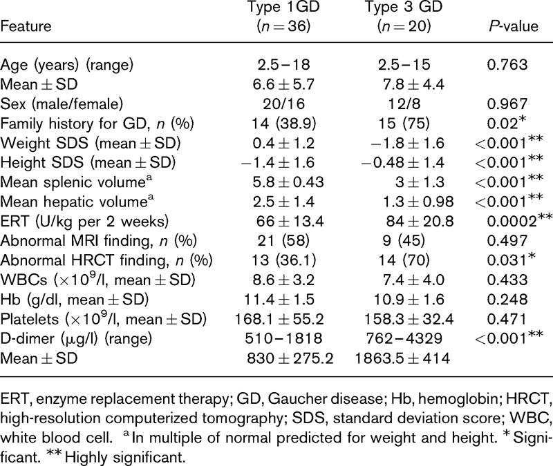
Forty‐eight adult patients with type I GD, 52 obligatory carriers, and 19 controls were enrolled to this study. The clinical characteristics of the GD patients are presented in Table I.

I  Characteristics of patients with type I Gaucher disease.
 No%
Total patients 48
Male 23 48
Ashkenazi Jewish origin 47 98
Mutations
 N370S homozygous 23 48
 Compound heterozygous (N370S/other) 25 52
Severity Score Index (SSI)
 Mild (1–10) 31 65
 Moderate (11–25) 16 33
 Severe (26–30) 1 2
 Splenectomy (total or partial) 17 35
 Medical therapy 36 75

Table 1

Effect of gaucher disease on platelet adhesion

Patients with type I GD had a significantly lower SC as compared to controls and obligatory carriers (Table II). The platelet counts of GD patients were not significantly different from controls; however they were significantly lower as compared to obligatory carriers. No differences in AS measurements were found between patients, obligatory carries and controls.


II  Laboratory characteristics of patients and obligatory carriers and healthy volunteers.

 
 PatientsControlsCarriers P‐value* P‐value† P‐value‡
Number (M:F) 48 (23:25) 19 (13:6) 52 (26:26)
Platelet count, ×109/l 193 (153–252) 222 (200–245) 249 (217–302) 0·06 0·003 0·06
Haematocrit, % 40·7 (37·3–44·1) 43 (40·5–45·8) 41·1 (37·2–45) 0·18 0·52 0·31
Surface coverage, % 4·6 (3·2–7·5) 8·7 (7. 6–10·3) 8·1 (6·5–9·4) 0·002 0·002 0·36
Average size, μm2 24·1 (20·1–33·9) 28·0 (24·0–34·0) 27·0 (24·0–37·0) 0·15 0·07 0·86
 

Data presented as median (interquartile range). M, male; F, female.


*P value for Gaucher patients compared to control.


P value for Gaucher patients compared to obligatory carriers.


P value for controls compared to obligatory carriers.

Table 2

Given that differences in platelet counts could potentially affect the difference observed in SC, 15 participants with platelet counts <160 × 109/l were excluded from the analysis in order to attain a median platelet count that was not significantly different between patients, obligatory carriers, and controls (P = 0·1). Also in this subgroups analysis, SC was significantly lower in patients, median (interquartile range), 5·9 (3·4–9), as compared to obligatory carriers, 8·1 (6·7–9·4) or controls, 8·8 (7·9–10·2; P = 0·001).

Correlation was found between SC and platelet counts in patients with type I GD (correlation coefficient 0·34, P  = 0·017, Spearman correlation test). No correlation was found with age and the SSI. No correlation was found between SC and platelet count in controls or in obligatory carriers. Representative photographs of platelet adhesion in a patient and a healthy control are shown (Fig 1).

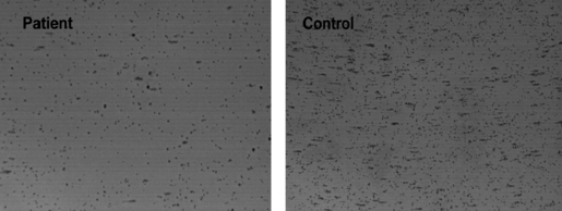
Figure 1  Reduced platelet adhesion in Gaucher disease. A representative platelet adhesion picture (IMPACT‐R, Cone and platelet analyser) of a healthy control (right): Surface coverage (SC) 9% Average size (AS) 27 μm2, and a patient with Type I Gaucher disease (left) SC 3·3%, AS 23 μm2.

Effect of ert and splenectomy on platelet adhesion

ERT‐treated patients had similar platelet counts, SC, and AS of aggregates compared to untreated patients (Table III). Splenectomized patients on ERT had significantly higher platelet counts and SC as compared to patients on ERT only. Only one splenectomized patient did not receive ERT and was excluded from this analysis.

III  Characteristics of patients with Gaucher type I with or without enzyme replacement therapy (ERT).
 No ERTERT onlyERT & splenectomy* P‐value† P‐value‡ P‐value§
Number 11 20 16
Age, years 39 (35–48) 40 (25–59) 48·5 (37–61) 0·82 0·16 0·12
Platelet count, ×109/l 154 (138–204) 169 (142–193) 262 (214–302) 0·99 0·002 <0·001
Haematocrit, % 40·6 (38·8–42·4) 39·7 (36·6–42·9) 41·3 (39·1–43·6) 0·44 0·89 0·58
Surface coverage, % 3·8 (1·9–7·5) 3·8 (2·4–5·3) 7·2 (5·8–9·3) 0·99 0·09 0·002
Average size, μm2 20·0 (17·1–25·1) 25·6 (22·5–35·8) 24·9 (22·3–31·7) 0·21 0·16 0·79
Mucosal bleeding 6 (54%) 7 (35%) 4 (25%) 0·29 0·15 0·7

Data presented as median (interquartile range). ERT, enzyme replacement therapy.


*One splenectomized patient who did not receive ERT was excluded from the analysis.


P value for patients without therapy compared to those who received ERT.


P value for patients without therapy compared to those who received ERT and underwent splenectomy.


§P value for patients who received ERT compared to who received ERT and underwent splenectomy.

Table 3

Although a trend of higher platelet count was found in splenectomized patients compared to controls (Fig 2A), the SC of those patients tended to be lower compared to controls (Fig 2B).


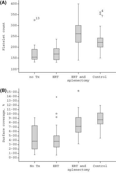
Figure 2  (A) Platelet counts in patients with Gaucher disease and controls. A box plot for the platelet count (×109/l) in untreated patients (no Rx, n = 11), patients receiving only ERT (ERT = 20), splenectomized patients also receiving ERT (ERT and splenectomy, n = 16), and controls (n = 19). A trend for higher platelet count in splenectomized patients on ERT compared to controls (P  = 0·066). (B) Surface coverage in patients with Gaucher disease and controls. A box plot curve for the surface coverage (% IMPACT‐R) in untreated patients (no Rx, n = 11), patients receiving only ERT (ERT n = 20), splenectomized patients receiving also ERT (ERT and splenectomy, n = 16) and controls (n = 19). A trend for lower surface coverage in splenectomized patients on ERT as compared to controls (P = 0·071).

No correlation was found between platelet counts and SC during analysis of the different treatment groups, i.e. untreated with intact spleen, ERT with intact spleen, and ERT with splenectomy.

Bleeding history in patients with gaucher disease

Twenty‐eight patients reported a previous history of bleeding episodes (58·3%, 95% CI 43·2%‐72·4%). Eleven patients (22·9%) reported only easy bruising. Mucosal bleeding was reported 17 patients (35·4%), including epistaxis, menorrhagia, gingival bleeding, rectal bleeding, and prolonged bleeding after cuts or surgery. A bleeding history was not obtained in one patient. A history of mucosal bleeding was not associated with the SSI, ERT, platelet counts, haematocrit level, and/or SC/AS measurements.

To improve the diagnostic value of the IMPACT‐R test as a potential predicative test for mucosal bleeding, the results of both the SC and the AS measurements were used to define an abnormal test. An abnormal IMPACT‐R test, (defined as an abnormal SC < 7% and/or AS < 23 μm2) was found in 32 patients (66·7%, 95% CI 51·6–79·6%) and was associated with history of mucosal bleeding. An abnormal IMPACT‐R test was found in 15/17 (88·2%, 95% CI 63·6–98·5%) of patients with mucosal bleeding as compared to 17/30 (56·6%, 95% CI 37·4–74·5%) of patients without mucosal bleeding (P = 0·037). The Odds Ratio (95% CI) for mucosal bleeding in GD patients with an abnormal IMPACT‐R test was 5·73 (1·1–29·6).

Mixing studies in patients and controls

Platelet adhesion in the IMPACT‐R system is influenced by platelets, RBCs, and plasma components (Shenkman , 2000; Peerschke , 2007). To test the differential role of each component in the observed reduced platelet adhesion in GD, we performed mixing studies in nine pairs of patients and controls. Patient PRP mixed with control RBC resulted in a lower SC compared to control PRP mixed with control RBC, mean (95% CI), 3·5% (1·5–5·5%) and 8·5% (7·2–9·8%), respectively (P < 0·001; Fig 3).


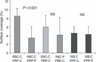
Figure 3  Mixing studies in patients and controls. RBC, red blood cell; PRP, platelet rich plasma; PPP, platelet poor plasma; WB, whole blood; C, control; P, patient; NS, not significant. Surface coverage (%) of reconstituted samples of patients and controls (n  = 9) as described in material and methods. Surface coverage was significantly reduced when RBC‐C were reconstituted with PRP‐P. No change in SC was observed when patient PPP or RBC was reconstituted with control blood.

Mixing of control PPP with control whole blood diluted the sample and reduced platelet adhesion, mean SC (95% CI), 4·54% (3·34–5·74%). However, mixing of patient PPP with control whole blood did not reduce platelet adhesion further: mean SC (95% CI), 4·29% (2·85–5·72%). Mixing of patient RBC with control PRP did not change platelet adhesion (for the whole group of patients, n = 8), mean SC (95% CI), 5·98% (3·7–8·26%; Fig 3). However, a splenectomized patients’ RBC mixed with normal PRP reduced SC as compared to autologous mixing of control RBC and control PRP (n = 5, P = 0·04). This phenomenon did not occur in the patients with intact spleens (n = 3, P = 0·4).

Platelet aggregation

Platelet aggregation in response to epinephrine, ADP, collagen and ristocetin was tested in 22 patients. Platelet counts, SC/AS measurements, and rate of abnormal IMPACT‐R test were not different in patients who were tested or not for platelet aggregation. Isolated abnormal platelet aggregation in response to epinephrine was observed in three (13%) patients (Kambayashi , 1996). Abnormal platelet aggregation in response to ADP and/or collagen was observed in five (22%) patients, all of whom had an abnormal IMPACT‐R test [positive predictive value of 100% (95% CI 89–100%)]. Aggregation in response to ristocetin was normal in all patients. Platelet aggregation defect alone was not associated with mucosal bleeding (P = 0·32).

Discussion

This study was designed to assess the platelet adhesion in patients with type I GD. Platelet adhesion was found to be lower in patients with GD compared to obligatory carriers and controls. Platelet adhesion was not affected by the use of ERT and was improved after splenectomy. A platelet adhesion defect, measured by the IMPACT‐R system, occurred in two‐thirds of GD patients and was associated with history of mucosal bleeding.

The IMPACT‐R system measures both the adhesion of platelets to the polystyrene surface (SC), as well as the aggregation of circulating platelets around adherent platelets associated with the release reaction (AS). The significantly lower SC in GD patients compared to controls suggests that a platelet adhesion defect is a major thrombocytopathy in patients with GD. This thrombocytopathy might be missed when testing platelet function only with the platelet aggregometer. Indeed, abnormal platelet aggregation was found in only one‐fifth of the patients in this study and others (Gillis , 1999; Giona , 2006).

The mechanism of reduced platelet adhesion in patients with type I GD is not clear. Based on mixing tests, reduced adhesion is an intrinsic platelets defect and not affected by RBCs or plasma of these patients. The increased plasma levels of glucocerebroside in patients with GD may affect platelet activation (Nilsson , 1982; Aerts & Hollak, 1997; Gousset , 2002). However, this mechanism could possibly explain platelet abnormalities only in untreated patients. Alternatively, extracts of platelets were found to be rich in an aggregated, activated form of the glucocerebrosidase (Yatziv , 1974), thus lack of the enzyme in GD might cause increased glucocerebroside in the platelets and affect platelet function.

In our study, patients receiving ERT did not have higher platelet counts or platelet adhesion compared to untreated patients. This is in contrast to previous studies where platelet counts and platelet aggregation were shown to be improved after starting ERT (Hollak , 1997; Gillis , 1999; Giona , 2006). By excluding patients with low platelet counts, we might have influenced the chance to find differences in platelet counts and platelet adhesion between ERT‐treated and untreated patients.

In contrast, splenectomy was associated both with improved platelet counts and platelet adhesion as measured by the IMPACT‐R system. The improved platelet adhesion found after splenectomy might be directly related to the higher platelet counts. Alternatively, splenomegaly has been suggested to cause chronic platelet activation that might lead to platelet exhaustion with reduced function (Humphries & Hess, 1994; Hollak , 1997).

The study inclusion criterion of a platelet count >130 × 109/l has resulted in a relatively high percentage of splenectomized patients (35%) in the era of disease‐specific ERT.

The trend to lower SC in splenectomized GD patients compared to controls, in the presence of a tendency for higher platelet counts suggests that the platelet adhesion defect we observed in GD patients is a true pathological finding and is not only a reflection of the low platelet counts. The observation of a lower SC in a subgroup analysis of patients with platelet counts >160 × 109/l, supports a similar conclusion.

Preliminary data from mixing studies indicate that RBC from GD patients with intact spleens may reduce the SC of normal platelets, as compared to RBC from splenectomized patients. These preliminary results suggest that the better platelet adhesion profile of splenectomized patients could also result from elimination of a yet unknown effect of splenomegaly on RBC physiology under high shear stress. Further research is needed to study the mechanism of the platelet adhesion defect in GD patients.

Of interest is the association of abnormal platelet function measured by the IMPACT‐R system with history of mucocutaneous bleeding. No similar association was reported with the more commonly performed platelet aggregation test. Studying whole blood platelet adhesion under high shear rate may be a more relevant parameter for correlation with bleeding symptoms. In previous studies, the IMPACT‐R method was shown to be effective in the assessment of platelet function in thrombocytopenic patients (Kenet , 1998) and in patients undergoing cardiac surgery (Gerrah , 2004). Recently, the IMPACT‐R method was shown to be a useful screening tool for bleeding disorders in children evaluated for bleeding tendency (Revel‐Vilk , 2009).

The diagnosis of bleeding risk is clinically important in preparing patients before surgery or delivery. The current recommendations in patients with GD include performing a complete history of bleeding tendency (including easy bruising and recurrent gum or nose bleeds, and heavy menstrual bleeds), a complete blood count, coagulation factors assays if the prothrombin time and/or partial thromboplastin time are abnormal, platelet aggregation tests, and von Willebrand factor levels and activity (Ioscovich , 2005; Zimran , 2005). Platelet adhesion studies are currently used in the Gaucher Clinic, Shaare Zedek Medical Centre, as part of the routine check of the haemostatic system before surgical procedures or delivery.

In conclusion, platelet adhesion defect is commonly found in type I GD patients and can explain some of the increased bleeding tendency in these patients.

Testing platelet adhesion should be considered before surgery, delivery or dental procedures in order to assess the need for platelet transfusions, antifibrinolytic agents or desmopressin (DDAVP) in the individual patient.

The first two authors contributed equally to the paper.


References

   (Exportez format texte tabulé)

  Aerts, J.M. & Hollak, C.E. (1997) Plasma and metabolic abnormalities in Gaucher’s disease. Baillieres Clinical Haematology, 10, 691–709.
  Berrebi, A., Malnick, S.D., Vorst, E.J. & Stein, D. (1992) High incidence of factor XI deficiency in Gaucher’s disease. American Journal of Hematology, 40, 153.
  Beutler, E. & Kuhl, W. (1970) Detection of the defect of Gaucher’s disease and its carrier state in peripheral‐blood leucocytes. Lancet, 1, 612–613.
  Beutler, E., Gelbart, T., Kuhl, W., Zimran, A. & West, C. (1992) Mutations in Jewish patients with Gaucher disease. Blood, 79, 1662–1666.
  Deghady, A., Marzouk, I., El‐Shayeb, A. & Wali, Y. (2006) Coagulation abnormalities in type 1 Gaucher disease in children. Pediatric Hematology and Oncology, 23, 411–417.
  Gerrah, R., Snir, E., Brill, A. & Varon, D. (2004) Platelet function changes as monitored by cone and plate(let) analyzer during beating heart surgery. The Heart Surgery Forum, 7, E191–E195.
  Gillis, S., Hyam, E., Abrahamov, A., Elstein, D. & Zimran, A. (1999) Platelet function abnormalities in Gaucher disease patients. American Journal of Hematology, 61, 103–106.
  Giona, F., Palumbo, G., Amendola, A., Santoro, C. & Mazzuconi, M.G. (2006) Platelet function and coagulation abnormalities in type 1 Gaucher disease patients: effects of enzyme replacement therapy (ERT). Journal of Thrombosis and Haemostasis, 4, 1831–1833.
  Gousset, K., Wolkers, W.F., Tsvetkova, N.M., Oliver, A.E., Field, C.L., Walker, N.J., Crowe, J.H. & Tablin, F. (2002) Evidence for a physiological role for membrane rafts in human platelets. Journal of Cellular Physiology, 190, 117–128.
  Granovsky‐Grisaru, S., Aboulafia, Y., Diamant, Y.Z., Horowitz, M., Abrahamov, A. & Zimran, A. (1995) Gynecologic and obstetric aspects of Gaucher’s disease: a survey of 53 patients. American Journal of Obstetrics and Gynecology, 172, 1284–1290.
  Hollak, C.E., Levi, M., Berends, F., Aerts, J.M. & van Oers, M.H. (1997) Coagulation abnormalities in type 1 Gaucher disease are due to low‐grade activation and can be partly restored by enzyme supplementation therapy. British Journal of Haematology, 96, 470–476.
  Humphries, J.E. & Hess, C.E. (1994) Gaucher’s disease and acquired coagulopathy. American Journal of Hematology, 45, 347–348.
  Ioscovich, A., Briskin, A., Abrahamov, A., Halpern, S., Zimran, A. & Elstein, D. (2005) Uncomplicated outcome after anesthesia for pediatric patients with Gaucher disease. Canadian Journal of Anaesthesia, 52, 845–847.
  Kambayashi, J., Shinoki, N., Nakamura, T., Ariyoshi, H., Kawasaki, T., Sakon, M. & Monden, M. (1996) Prevalence of impaired responsiveness to epinephrine in platelets among Japanese. Thrombosis Research, 81, 85–90.
  Katz, K., Tamary, H., Lahav, J., Soudry, M. & Cohen, I.J. (1999) Increased operative bleeding during orthopaedic surgery in patients with type I Gaucher disease and bone involvement. Bulletin/Hospital for Joint Diseases, 58, 188–190.
  Kelsey, H., Christopoulos, C., Gray, A.A. & Machin, S.J. (1994) Acquired pseudo‐pseudo Bernard‐Soulier syndrome complicating Gaucher’s disease. Journal of Clinical Pathology, 47, 162–165.
  Kenet, G., Lubetsky, A., Shenkman, B., Tamarin, I., Dardik, R., Rechavi, G., Barzilai, A., Martinowitz, U., Savion, N. & Varon, D. (1998) Cone and platelet analyser (CPA): a new test for the prediction of bleeding among thrombocytopenic patients. British Journal of Haematology, 101, 255–259.
  Larsen, E.C., Connolly, S.A. & Rosenberg, A.E. (2003) Case records of the Massachusetts General Hospital. Weekly clinicopathological exercises. Case 20‐2003. A nine‐year‐old girl with hepatosplenomegaly and pain in the thigh. New England Journal of Medicine, 348, 2669–2677.
  Nilsson, O., Hakansson, G., Dreborg, S., Groth, C.G. & Svennerholm, L. (1982) Increased cerebroside concentration in plasma and erythrocytes in Gaucher disease: significant differences between type I and type III. Clinical Genetics, 22, 274–279.
  Peerschke, E.I., Silver, R.T., Weksler, B.B., Yin, W., Bernhardt, B. & Varon, D. (2007) Examination of platelet function in whole blood under dynamic flow conditions with the cone and plate(let) analyzer: effect of erythrocytosis and thrombocytosis. American Journal of Clinical Pathology, 127, 422–428.
  Revel‐Vilk, S., Varon, D., Shai, E., Agmon, Y., Hyam, E., Daas, N., Miskin, H. & Weintraub, M. (2009) Evaluation of children with a suspected bleeding disorder applying the Impact‐R [Cone and Plate(let) Analyzer]. Journal of Thrombosis and Haemostasis, 7, 1990–1996.
  Seligsohn, U., Zitman, D., Many, A. & Klibansky, C. (1976) Coexistence of factor XI (plasma thromboplastin antecedent) deficiency and Gaucher’s disease. Israel Journal of Medical Sciences, 12, 1448–1452.
  Shenkman, B., Savion, N., Dardik, R., Tamarin, I. & Varon, D. (2000) Testing of platelet deposition on polystyrene surface under flow conditions by the cone and plate(let) analyzer: role of platelet activation, fibrinogen and von Willebrand factor. Thrombosis Research, 99, 353–361.
  Varon, D., Dardik, R., Shenkman, B., Kotev‐Emeth, S., Farzame, N., Tamarin, I. & Savion, N. (1997) A new method for quantitative analysis of whole blood platelet interaction with extracellular matrix under flow conditions. Thrombosis Research, 85, 283–294.
  Wright, S. (1992) Adjusted P‐values for simultaneous inference. Biometrics, 48, 1005–1013.
  Yatziv, S., White, M. & Eldor, A. (1974) Lysosomal enzyme activities in platelets of normal individuals and of patients with Gaucher’s disease. Thrombosis et diathesis haemorrhagica, 32, 665–669.
  Zimran, A., Sorge, J., Gross, E., Kubitz, M., West, C. & Beutler, E. (1989) Prediction of severity of Gaucher’s disease by identification of mutations at DNA level. Lancet, 2, 349–352.
  Zimran, A., Altarescu, G., Rudensky, B., Abrahamov, A. & Elstein, D. (2005) Survey of hematological aspects of Gaucher disease. Hematology, 10, 151–156.


British Journal of Haematology 2011; 153(3): 372-8


lierre_fleur445


13 mai 2011

D-dimères et atteintes osseuses et pulmonaires dans la maladie de Gaucher (D-dimer assay in Egyptian patients with Gaucher disea

Article classé dans la catégorie : "Examens biologiques, IRM,...".

Vous trouverez des liens utiles à la suite de la rubrique "CATEGORIE".

Ghislaine SURREL

maladies-lysosomales-subscribe@yahoogroupes.fr

D-dimères et atteintes osseuses et pulmonaires dans la maladie de Gaucher Dans la maladie de Gaucher, les atteintes osseuses et pulmonaires constituent 2 causes majeures de morbidité. Pour évaluer le caractère prédictif potentiel du taux de D-dimères (indicateur fiable d’une thrombose microvasculaire active), une étude égyptienne a inclus 56 patients présentant une maladie de Gaucher (36 types 1 et 20 types 3) et 30 témoins bien portants. Le taux de D-dimères était significativement plus élevé chez tous les patients par rapport aux témoins et chez les patients avec une maladie de type 3 par rapport aux patients avec une maladie de type 1. Une proportion plus importante de patients avec un type 3 présentait une atteinte pulmonaire et une proportion plus importante de patients avec un type 1 présentait une atteinte osseuse. Le taux de D-dimères était significativement plus élevé en cas d’anomalies des os longs à l’IRM et en cas d’aspect en verre dépoli au scanner thoracique. Les patients splénectomisés présentaient un taux plus élevés de D-dimères que ceux qui ne l’étaient pas. Selon les auteurs ces résultats montrent que le taux de D-dimères est significativement élevé dans la maladie de Gaucher, en particulier dans le type 3, et suggèrent qu’il représente un marqueur prédictif potentiel d’atteinte des os et des poumons utilisable au cours de la surveillance de la réponse au traitement.

D-dimer assay in Egyptian patients with Gaucher disease: correlation with bone and lung involvement

[13-05-2011]

Sherif, Eman Ma; Tantawy, Azza AGa; Adly, Amira AMa; Kader, Hossam Ab; Ismail, Eman ARc

aDepartment of Pediatrics, Egypt, bDepartment of Radiology, Egypt, cDepartment of Clinical Pathology, Faculty of Medicine, Ain Shams University, Cairo, Egypt

Received 20 August, 2010, Revised 17 October, 2010, Accepted 27 October, 2010

Correspondence to Eman A.R. Ismail, MD, 5 Nageb Mahfoz Street, Agouza, 12654 Giza, Egypt Fax: +202 3337 5435; e-mail: eman.ismail_70@yahoo.com

Abstract

Gaucher disease is the most frequent lysosomal storage disorder. Bone and lung involvement are two major causes of morbidity in this disease. D-dimer is a reliable indicator of active microvascular thrombosis, even in patients without overt hypercoagulation. This study aimed to assess D-dimer levels in Gaucher disease, correlating this marker to clinical characteristics and radiological parameters to investigate its role as a potential predictor for the occurrence and severity of skeletal and pulmonary manifestations. The study population consisted of 56 Egyptian patients with Gaucher disease, 36 had type 1 Gaucher disease (64.3%) and 20 had type 3 Gaucher disease (35.7%). Thirty healthy individuals were enrolled as a control group. D-dimer levels were significantly higher in all patients with Gaucher disease compared with controls (P < 0.001). Patients with type 3 showed significantly higher D-dimer concentrations compared with type 1 (P < 0.001). Pulmonary involvement was present in a significant proportion among type 3 Gaucher patients (P < 0.05), whereas bone changes were present in a higher percentage in type 1 compared with type 3 Gaucher patients. D-dimers were significantly higher in patients with abnormal MRI findings of the long bones and in those with ground glass appearance on high-resolution computerized tomography of the chest compared with patients with normal radiology (P < 0.001). Splenectomized patients displayed significantly higher D-dimer levels compared with nonsplenectomized patients (P < 0.001). Our results suggest that D-dimer is significantly elevated in Gaucher disease, particularly type 3, and may be considered as a potential marker of risk prediction of bone and lung involvement that could be used to monitor treatment response.

Introduction

The lysosomal storage diseases have a cumulative incidence of 1 in 5000 live births [1]. Gaucher disease predominates among this group, having a frequency of 1 in 40 000 in the United States [2,3]. Gaucher disease results from an autosomal recessive deficiency of the lysosomal enzyme acid [beta]-glucosidase [glucocerebrosidase (GBA)], which is responsible for hydrolysis of glucocerebroside [glucosylceramide (GLC)] [4]. Mutations in the GBA gene result in Gaucher disease and many of these mutations are missense alterations that may cause misfolding, decreased stability and/or mistrafficking of this lysosomal protein [5]. Absent or reduced enzymatic activity leads to accumulation of GLC in various cells of the macrophage–monocyte system (Gaucher cells) [6,7]. Classically, the pathophysiology of Gaucher disease has been attributed to the amount, location and rate of accumulation of the stored material [8,9]. The manifestations of Gaucher disease occurring due to accumulation of Gaucher cells in three main anatomical compartments, namely the osseous skeleton, the bone marrow and visceral organs [4,10].

As with most genetic diseases, the signs and symptoms of Gaucher disease present along a continuum, ranging from the lethal neonatal form to the asymptomatic form [11]. Currently, Gaucher disease is classified into three clinical forms: nonneuropathic (type 1), acute neuropathic (type 2) and chronic neuropathic (type 3) [3,10]. Patients with type 1 disease may present at any age with hepatosplenomegaly, anemia, thrombocytopenia, often skeletal involvement (such as avascular necrosis of the large joints or pathological fractures) or lung disease. Age and mode of presentation as well as the eventual clinical course are highly variable, yet phenotypic heterogeneity can be attributed only in part to specific mutations [9,12].

Bone disease usually designates the advanced stages of Gaucher disease, but susceptibility to fractures and avascular necrosis can be the first sign of Gaucher disease in otherwise asymptomatic patients [13]. The skeletal aspects of the disease have a much greater impact on patients' quality of life than the hematological and visceral aspects. Moreover, skeletal manifestations are commonly seen in patients with normal hematology [14]. Therefore, it is important for physicians not to focus only on hematological and visceral complications on the expense of skeletal involvement when assessing the response to enzyme replacement therapy (ERT) [15,16]. Nearly, all patients with Gaucher disease have radiological evidence of skeletal involvement including Erlenmeyer flask deformity, osteopenia, osteosclerosis, osteonecrosis, fractures and bone marrow infiltration. Skeletal involvement follows three basic processes: focal disease (irreversible lesions such as osteonecrosis and osteosclerosis), local disease (reversible abnormalities adjacent to heavily involved marrow such as cortical thinning and long bone deformity) and generalized osteopenia [17]. For more accurate assessment of bone disease in adults and children, it is desirable to conduct MRI studies at centers with radiologists experienced in evaluating patients with Gaucher disease [18].

Symptomatic lung involvement may be common at presentation, and may progress over the course of the disease to pulmonary hypertension in some patients, particularly in type 3 Gaucher disease [13]. Chest radiographs may demonstrate reticulonodular changes, and on high-resolution computerized tomography (HRCT), a range of abnormal patterns including widespread ground glass opacification may be present [19,20]. 

Many factors may trigger or aggravate symptoms and signs of the disease including environmental or acquired conditions, such as viral infections or pregnancy. In addition, other concurrent genetic defects such as partial deficiency of coagulation factors aggravate the tendency to bleeding and may contribute to the variability of clinical manifestations in Gaucher patients. These modifiers may be helpful in predicting risk of developing bone and/or lung disease [3,10,21]. A logical theory to explain the incidence of avascular bone necrosis and pulmonary hypertension in Gaucher disease was an inherited predilection for hypercoagulability [9,22]. Gaucher disease has been the playground to develop new therapeutic interventions such as ERT and substrate-reduction therapy. The availability of these costly therapies has stimulated research regarding suitable biomarkers to monitor onset and progression of disease, as well as the efficacy of therapeutic intervention [23].

D-dimer assays are ordered, along with laboratory tests that help to rule out, diagnose and monitor conditions associated with hypercoagulability [24,25]. The possibility of correlation between avascular bone necrosis and pulmonary hypertension in Gaucher patients with D-dimer levels was considered because it would implicate involvement of microthrombosis, without expecting a quantitative correlation with hypercoagulability per se [9]. However, there have been few studies that evaluated the impact of this reliable indicator of coagulation on the development and severity of skeletal and pulmonary manifestations in Gaucher disease.

Therefore, this study aimed to assess D-dimer levels in Egyptian patients with Gaucher disease as a potential predictor for the occurrence and severity of bone and lung involvement correlating this marker to clinical characteristics and radiological parameters.

Patients and methods

This cross-sectional study was conducted on 56 consecutive patients with Gaucher disease attending the Hematology/Oncology Clinic in Pediatric Hospital, Ain Shams University, from June 2009 to May 2010. They were 33 men and 23 women, with a male to female ratio of 1.4: 1. The mean age at diagnosis in the study population as a whole was 6.0 ± 3.9 years (range, 2.5–13 years). Control samples (peripheral blood) were obtained from 30 healthy children and adolescents with no history of inborn errors of metabolism in their first-degree and second-degree relatives. Twelve were men and 18 were women (male to female ratio, 1: 1.5). The mean age of the control population was 7.4 ± 4.24 years (range, 3–15 years). A written consent was obtained from the guardian of each case and control before participation in the study. The study was approved from the local ethical committee.

All patients were subjected to full medical history with special emphasis on bone manifestations (pains or fractures), hematological manifestations (pallor, bleeding and history of blood transfusion), pulmonary manifestations (cough, dyspnea and recurrent chest infections) and neurological symptoms (squint, convulsions, bulber manifestations, trismus and manifestations suggestive of cerebellar affection). Thorough clinical examination was performed laying stress on anthropometric measures, abdominal examination with assessment of hepatic and splenic size and full neurological examination including cranial nerves, motor and sensory systems. Radiological investigations included plain radiograph on long bones, chest radiograph, abdominal ultrasonography to assess the volume of liver and spleen in cubic centimeter (calculated as multiples of normal sizes predicted for body weight) [15] and MRI on pelvis and femur, as well as HRCT on both lung fields.

Laboratory investigations for Gaucher patients included complete blood count, examination of peripheral blood-stained smears, bone marrow aspiration and examination of stained smears and coagulation profile, as well as liver and renal function tests. GBA enzyme activity was assessed in peripheral blood leukocytes and plasma chitotriosidase was measured at diagnosis and for follow-up of the response of ERT as a sensitive indicator of dose effects. Molecular analysis of the acid GBA gene was performed for 18 patients.

Diagnostic criteria

Diagnosis of Gaucher disease was based on the presence of hepatosplenomegaly, bone involvement, hematological manifestations and developmental delay with or without neurological manifestations in the form of occulomotor abnormalities and/or convulsions. This was confirmed by laboratory findings of Gaucher cells in bone marrow aspirate and biopsy, low GBA activity [4] and high plasma level of chitotriosidase activity [26,27].

The clinical and laboratory characteristics of the studied Gaucher patients are summarized in Table 1. The studied patients were divided into two groups: 

Table 1 Clinicopathological characteristics of the studied Gaucher patients

Group I included 36 patients with type 1 Gaucher disease (64.3%). They were 20 men and 16 women with a male to female ratio of 1.3: 1. Their ages at the time of the study ranged from 2.5 to 18 years with a mean of 6.6 ± 5.7 years. Type 1 patients presented initially with hepatosplenomegaly, bony manifestations, anemia and thrombocytopenia. At time of the study, 19 of 36 type 1 patients had anemia and 15 had thrombocytopenia as well as organomegaly. The duration of treatment ranged from 1 to 8 years with a mean of 2.9 ± 2.4 years. They were under ERT in a dose ranging from 60 to 90 U/kg per 2 weeks with a mean of 66 ± 13.4 U/kg per 2 weeks.

 Group II included 20 patients with type 3 Gaucher disease (35.7%). They were 12 men and eight women (male to female ratio, 1.5: 1). Their ages at the time of the study ranged from 2.5 to 15 years with a mean of 7.8 ± 4.4 years. Type 3 patients presented with neurological manifestations in the form of ophthalmoplegia, neck stiffness, dysphagia, convulsions and trismus, in addition to visceral and hematological manifestations. The duration of treatment ranged from 1.5 to 8 years with a mean of 6.6 ± 5.7 years. They were under ERT in a dose ranging from 60 to 120 U/kg per 2 weeks with a mean of 84 ± 20.8 U/kg per 2 weeks.

 None of the studied patients had advanced liver disease, active inflammatory conditions or were on anticoagulant therapy at time of study. Fourteen patients (35%) were splenectomized prior to the onset of ERT. All patients were under regular ERT [28,29] for a period of 1–8 years starting from January 1999. ERT was first given using the placenta-derived aglucerase (Ceredase; Genzyme Corporation, Cambridge, Massachusetts, USA) at low dose regimen (15 U/kg per 2 weeks). The recombinant enzyme imiglucerase (Cerezyme; Genzyme Corporation) was applied in March 1999 in a dose of 20–30 U/kg per month, divided into four equal weekly doses. In July 1999, Cerezyme was administered in a dose of 60 U/kg per 2 weeks. Six patients with type 3 received high doses of 120 U/kg per 2 weeks, one of them had progressive neurological disease and the others suffered severe pulmonary involvement.

 Blood sampling and assay of d-dimer

 As part for routine work-up for Gaucher disease, peripheral blood samples were collected on 3.2% buffered sodium citrate at a ratio of nine parts blood to one part anticoagulant (1: 10 ratio) and centrifuged at 1600 g for 15 min. D-dimer concentrations were determined in platelet-poor plasma using Tina-quant assay (Roche Diagnostics, Mannheim, Germany) on a Hitachi 917 analyzer (Roche Diagnostics). Tina-quant D-dimer is an immunoturbidimetric assay in which a beam of monochromatic light is passed through the suspension of latex microparticles coated by covalent bonding with monoclonal antibodies specific for D-dimer. The wavelength of the light is greater than the diameter of the latex microparticles and thus the solution of latex microparticles only slightly absorbs the light. In the presence of D-dimer, the particles aggregate and turbidity increases. The increase in scattered light is proportional to the amount of D-dimer in the test sample. The latex particles are coated with a monoclonal antibody that reacts with fibrin D-dimer or fragment D of fibrin. The antibody has no cross-reactivity with fibrinogen. This allows quantitative determination of D-dimer in human plasma. Normal limit of the assay is 500 µg/l. The test has a lower detectable limit of 40 µg/l [25,30–32].

 Statistical analysis

 The processing of data was computed using statistical package for social science (SPSS), version 14 IBM compatible PC (SPSS Inc., Chicago, Illinois, USA). Data were described in the form of number and percentage, and range and mean ± SD. In order to compare quantitative variables between two groups, both the Student's t-test and the nonparametric Mann–Whitney U-test were applied. A [chi]2-test was used to compare qualitative variables. A P-value of 0.05 or less and 0.01 or less was considered significant and highly significant, respectively, in all analyses.

Results

 Clinical, hematological and radiological features of gaucher patients

 Type 1 Gaucher disease was found in 64.3% of patients, and type 3 in 35.7%. Molecular analysis of the acid GBA gene in 18 patients revealed homozygosity for L444P in 11 patients (all were type 3), whereas six type 1 Gaucher patients showed mutation of N370S and only one patient with type 1 Gaucher disease displayed the uncommon homozygous D409H mutation.

 As shown in Table 1, age and sex did not differ significantly between patients in both groups, although positive family history for Gaucher disease was significantly present in type 3 compared with type 1 Gaucher patients (P < 0.05). Underweight was frequently observed in type 3 patients, whereas short stature was frequently observed in type 1 (P < 0.001). Mean hepatic and splenic volumes reported in multiples of normal size predicted for body weight in type 1 Gaucher disease were significantly higher when compared with those of type 3 patients (P < 0.001). White blood cell count, hemoglobin levels and platelet counts did not show any significant difference between both study groups of Gaucher disease (P > 0.05). Type 3 Gaucher patients received significantly higher doses of ERT compared with type 1 (P < 0.001).

 As regards radiological findings (Table 1), in type 1 Gaucher disease, 21 patients (58%) had Erlenmeyer flask-shaped deformity in radiograph of long bones, as well as bone marrow expansion with cortical thinning in MRI. Moreover, 13 patients (36.1%) had ground glass appearance on HRCT chest. In type 3 Gaucher disease, nine patients (45%) had Erlenmeyer flask-shaped deformity in radiograph and bone marrow expansion with cortical thinning in MRI (Fig. 1). Fourteen patients (70%) had ground glass appearance in HRCT chest (eight of them had interlobular thickening). Pulmonary involvement shown by HRCT results (Figs 2 and 3) was present in a significantly higher percentage among type 3 Gaucher patients compared with type 1 (P < 0.05). Although bone changes represented by MRI findings were present in a higher percentage in type 1 compared with type 3 Gaucher disease, yet the difference did not reach statistical significance (P > 0.05). It was noticed that the studied Gaucher patients under ERT in a dosage of 60 U/kg per 2 weeks suffered from clinically and radiologically evident pulmonary symptoms confirmed by HRCT findings. However, when these patients received higher doses of ERT ranging from 90 to 120 U/kg per 2 weeks, a remarkable improvement in their clinical manifestations was observed. The frequency and severity of chest infections that required hospitalization were markedly decreased. There was decreased dyspnoea, clubbing and cyanosis in some patients, although on radiology, lung pathology was not normalized. Others showed improved respiratory compliance, with a significant improvement of the radiological findings.

 D-dimer levels in control population and gaucher patients

 D-dimer was negative (<500 µg/l) in controls with a range of 77.8–410 µg/l and a mean value of 215.7 ± 32.5 µg/l. All patients with Gaucher disease had significantly elevated D-dimer levels, with a range of 510–4329 µg/l and a mean of 2018 ± 368 µg/l at time of the study (P < 0.001). Patients with type 1 and type 3 Gaucher disease displayed significantly high D-dimer concentrations than controls when compared separately (P < 0.001) (Fig. 4). Moreover, D-dimer levels were significantly higher in type 3 patients compared with type 1 (mean, 1863.5 ± 414 vs. 830 ± 275.2 µg/l; P < 0.001) (Table 1 and Fig. 4).

 D-dimer analysis results in relation to clinical and radiological data

 Analysis of D-dimer in relation to clinical and radiological data of Gaucher patients (Table 2) revealed significantly higher levels in patients with abnormal MRI findings of long bones compared with those with normal MRI study (mean, 1563.6 ± 357 vs. 810.7 ± 250 µg/l; P < 0.001). In addition, Gaucher patients with pulmonary involvement on HRCT chest had higher D-dimer concentrations compared with those with normal radiological study (mean, 1980 ± 400 vs. 990 ± 205 µg/l; P < 0.001). Splenectomized patients displayed significantly higher D-dimer levels compared with nonsplenectomized patients (mean, 1670 ± 326 vs. 746 ± 187 µg/l; P < 0.001).


Table 2 Clinical and radiological features of the studied Gaucher patients according to D-dimer levels

Discussion

Studies of genotype–phenotype correlations in Gaucher disease, the most common sphingolipidosis, revealed significant genotypic heterogeneity among clinically similar patients and vastly different phenotypes among patients with the same mutations [11]. The incidence of the subacute neuronopathic (type 3) form is less than one in 100 000 patients. The distribution of type 3 Gaucher disease is panethnic, but this form predominates in some geographic regions such as northern Sweden [33,34]. In the present study, type 3 Gaucher disease was found in 35.7% of patients. This could be explained by the higher prevalence of the genotype L444P among this ethnic group as it was reported that N370S homozygotes generally present with a less severe phenotype, whereas L444P and D409H homozygosity confers neurologic involvement [2,4,35]. This is in contrast to Ashkenazi Jewish population in which the genotype N370S is predominant, resulting in higher prevalence of type 1 Gaucher disease with mild clinical manifestations [13]. Moreover, some genetic mutations are unique to individual groups or families. For instance, D409H/D409H causes calcification of the heart valves and occulomotor apraxia, but without visceral affection occurring uniquely in Jenin Arabs, Japanese and Spanish patients [36,37].

 Failure to thrive and underweight were prominent features in the studied patients, especially in type 3 Gaucher disease. In line with our results, Zimran et al. [38] noticed that poor weight gain and growth retardation in those patients occurred likely due to the excessive metabolic burden from accumulation of undegraded substrate and GLC in tissues, together with the chronic nature of the disease and anemia that accounts for the chronic fatigue reported by many patients. The low-grade, smoldering and subclinical internal inflammation in individuals with Gaucher disease is accompanied by an increased degree of erythrocyte and leukocyte adhesiveness/aggregation. These findings might have rheological consequences in terms of microcirculatory slow flow and tissue hypoxemia. An additional factor in type 3 Gaucher patients is pseudobulbar palsy resulting in dysphagia, regurgitation and recurrent aspiration pneumonia [39]. Similarly, Khalifa et al. [40] reported that patients with Gaucher disease were more susceptible to recurrent infection, which is a main cause of debilitation. The tendency toward infection in Gaucher disease patients was attributed to their postsplenectomy state. Furthermore, Orvisky et al. [8] noticed resumption of normal growth in children after receiving ERT.

As previously reported, the estimated life expectancy for patients with type 1 Gaucher disease was mildly decreased than the reference population being 9 years less [41]. Disease progression varies in type 1 Gaucher disease and survival may be normal depending on the severity of complications. On the contrary, evolution is rapid in type 2 Gaucher disease leading to death within the first 2 years of life, usually because of lung failure, whereas type 3 Gaucher disease patients often survive to the second or third decades of life [2,42]. In this study, the significantly higher doses of ERT received by patients with type 3 Gaucher disease compared with type 1 were in concordance to the report of Vellodi et al. [43], who stated that there is clear evidence that ERT improves systemic involvement in nonneuronopathic as well as neuronopathic Gaucher disease enhancing quality of life. In addition, ERT has reversed, stabilized or slowed the progression of neurological involvement in some patients and is considered the gold standard treatment for Gaucher disease type 1 and type 3 patients [27,43,44]. High dose provides a faster clinical response and should be considered for patients with more aggressive disease [45].

 Despite some general genotype–phenotype correlations, disease severity and clinical outcomes cannot be predicted on the basis of genotype [13,46]. Genetic modifiers may play an important role in determining the eventual Gaucher phenotype [39]. Identification of biochemical markers characteristic of pathology may be of use in predicting the progression of the disease state [23,34,47]. There are no predictive tests to ascertain patients at risk for bone and lung involvement in Gaucher disease, which are slow to respond to ERT [4,9]. As reported by Deghady et al. [48], bleeding tendency in Gaucher disease may not always be related to absolute platelet counts, but may be influenced by coagulation factor deficiencies or abnormal platelet function. Acquired coagulation factor deficiencies have been demonstrated in Gaucher disease. The mechanism involved is unclear, but may include low-grade disseminated intravascular coagulation or sequestration of coagulation factors by substrate-loaded cells [10].

 Quantitative D-dimer determination has become routine practice in patients evaluated for the presence of deep venous thrombosis or pulmonary emboli [25,49]. In this study, plasma fibrin D-dimer was assayed in Gaucher disease using a new quantitative latex test for cross-linked fibrin degradation products, involving a specific monoclonal antibody. This method has been proven to be rapid, sensitive and reproducible [30].

The relevance of the determination of D-dimer in Gaucher disease was evaluated and high plasma D-dimer levels were found among patients with Gaucher disease, especially type 3. In addition, a significant elevation was observed in those with bone changes and pulmonary involvement as reflected by radiological findings. In agreement with our results, Shitrit et al. [9] stated that the levels of elevated of D-dimers were related to the presence of pulmonary infiltration, by Gaucher cells, as well as bone marrow infiltration. They implied that microthrombi may be part of the pathogenesis for avascular necrosis as well as pulmonary hypertension in patients with Gaucher disease. The significant correlation of D-dimer levels with treatment may be an indirect marker of bone and/or lung in Gaucher disease [9]. Additionally, Boot et al. [33] and Maire et al. [34] reported that the elevation of the serum concentration of several serologic markers (e.g. D-dimer, CCL18/PARC, CD163) in persons with Gaucher disease is considered a possible surrogate indicator of disease burden that could be used in monitoring treatment response.

 Moreover, it was reported that a normal MRI study in Gaucher patients with elevated D-dimer levels does not rule out subsequent development of bony changes in those patients, especially if splenectomized. Therefore, serial follow-up is required and the rising titer of D-dimer may determine or indicate the proper timing of the next MRI study [20].

 Strikingly, higher D-dimer concentrations were found among splenectomized patients with Gaucher disease. These results were in line with those obtained by Ashkenazi et al. [50], who observed that patients with Gaucher disease usually start to suffer from severe bony manifestations and pulmonary hypertension after splenectomy. In a preliminary study, Tunaci et al. [51] reported that asplenia was strongly associated with life-threatening forms of pulmonary hypertension in Gaucher disease, as removal of the spleen, which is the primary reservoir of storage cells, promotes migration of the mononuclear phagocyte system toward other tissue macrophage pools (lung and bone). Recently, Mistry et al. [52] observed a higher risk of avascular necrosis was observed among patients who had previously undergone splenectomy.

 Bone involvement was evident clinically and radiologically in the studied patients with type 1 Gaucher disease while pulmonary involvement reflected by HRCT findings was significantly found among type 3 Gaucher disease compared to type 1. This finding could be explained by the significantly elevated D-dimer levels encountered in type 3 Gaucher patients compared to type 1 Gaucher disease which is a less severe form. In this context, El-Beshlawy et al. [46] reported that bone manifestations were the most common presenting symptoms in Egyptian patients with type 1 Gaucher disease as evidenced by MRI long bones and ERT was effective in ameliorating radiological manifestations of skeletal disease and achieving complete remission of bone pain but it required a long period of treatment. Moreover, Jmoudiak and Futerman [13] reported that lung involvement including interstitial lung disease and pulmonary hypertension was found in a small number of patients with type 1 Gaucher disease. Additionally, Miller et al. [53] reported a higher frequency of pulmonary involvement in type 3 Gaucher disease compared to type 1. On the other hand, few patients develop pulmonary hypertension on ERT [54]. Mistry et al. [22] also revealed a remarkable predisposition for pulmonary hypertension in type 1 Gaucher disease. Progression to severe, life-threatening pulmonary hypertension occurs in the presence of additional genetic factors (non-N370S GBA mutation, positive family history and angiotensin converting enzyme I gene polymorphism) and epigenetic modifiers (i.e. asplenia, female sex) [22]. Therefore, routine echocardiographic monitoring of all treated and untreated patients with type 1 Gaucher disease was recommended and consideration of treatment withdrawal was suggested if the TI gradient progresses to more than 30 mmHg [54].

 In the present analysis, the remarkable improvement occurred in pulmonary manifestations with higher doses of ERT confirmed the previous reports by Elstein et al. [54] and Ashkenazi et al. [50], who noticed that the lung and bone are less accessible compartments to ERT, so that a low-dose regimen may not be sufficient to overcome a threshold for Gaucher cell infiltration and the dose must be increased to allow an adequate enzyme concentration at the site of the pathology, especially if there are fibrotic or necrotic regions. Moreover, Goitein et al. [55] reported that in patients with Gaucher disease and symptomatic lung involvement, there is great heterogeneity in presentation and response to ERT. Clinically, some benefited significantly from ERT, but in contrast to the dramatic reduction in organomegaly, there was no normalization in pulmonary function or lung architecture. These findings emphasized the importance of optimal dose of ERT, avoidance of splenectomy and vigorous combination therapy with vasodilators, as appropriate for the best outcomes in Gaucher patients [16,29,43].

 In conclusion, elevated D-dimer levels in Gaucher patients can be used as a predictor for the development of skeletal and pulmonary complications and may suggest that the occurrence of these pathological changes is induced initially as microthrombi. Thereafter, it would be interesting to apply the fully quantitative automated D-dimer assay in a prospective manner to ‘borderline’ Gaucher patients with persistent dyspnea, bronchorrhea and recurrent pneumonopathy, to ascertain whether they have merely incipient pulmonary changes and follow-up them for the possibility of developing progressive pulmonary hypertension. Further studies are recommended to indicate whether a predictive cut-off level of D-dimer in Gaucher disease can be described.

References

[1.] Meikle PJ, Hopwood JJ. Lysosomal storage disorders: emerging therapeutic options require early diagnosis. Eur J Pediatr 2003; 162(Suppl. 1):S34–S37.

[2.] Martins AM, Valadares ER, Porta G, Coelho J, Filho JS, Pianovski MAD, et al. Recommendations on the diagnosis, treatment, and monitoring of Gaucher disease. J Pediatr 2009; 155(Suppl. 2):S10–S18.

[3.] Mehta A. Epidemiology and natural history of Gaucher's disease. Eur J Intern Med 2006; 17:S2–S5.

[4.] Beutler E, Grabowski GA. Gaucher disease. In: Scriver CR, Valle D, Beudet A, Sly WS, editors. The metabolic and molecular bases of inherited diseases. Volume III. New York: McGraw-Hill; 2001. pp. 3635–3668.

[5.] Urban DJ, Zheng W, Goker-Alpan O, Jadhav A, Lamarca ME, Inglese J, et al. Optimization and validation of two miniaturized glucocerebrosidase enzyme assays for high throughput screening. Comb Chem High Throughput Screen 2008; 11:817–824.

[6.] Grabowski GA. Gaucher disease: lessons from a decade of therapy. J Pediatr 2004; 144:S15–S19.

[7.] Mignot C, Doummar D, Maire I, de Villemeur TB. Type 2 Gaucher disease: 15 new cases and review of the literature. Brain Dev 2006; 28:39–48.

[8.] Orvisky E, Park JK, LaMarca ME, Ginns EI, Martin BM, Tayebi N, et al. Glucosylsphingosine accumulation in tissues from patients with Gaucher disease: correlation with phenotype and genotype. Mol Genet Metab 2002; 76:262–270.

[9.] Shitrit D, Rudensky B, Zimran A, Elstein D. D-Dimer assay in Gaucher disease: correlation with severity of bone and lung involvement. Am J Hematol 2003; 73:236–239.

[10.] Hughes D, Cappellini MD, Berger M, Droogenbroeck JV, de Fost M, Janic D, et al. Recommendations for the management of the haematological and onco-haematological aspects of Gaucher disease. Br J Haematol 2007; 138:676–686.

[11.] Sidransky E. Gaucher disease: complexity in a ‘simple’ disorder. Mol Genet Metab 2004; 83:6–15.

[12.] Mistry P, Germain DP. Phenotype variations in Gaucher disease. Rev Med Interne 2006; 27(Suppl. 1):S3–S10.

[13.] Jmoudiak M, Futerman AH. Gaucher disease: pathological mechanisms and modern management. Br J Haematol 2005; 129:178–188.

[14.] Poll LW, Maas M, Terk MR, Roca-Espiau M, Bembi B, Ciana G, et al. Response of Gaucher bone disease to enzyme replacement therapy. Br J Radiol 2002; 75(Suppl. 1):A25–A36.

[15.] Elstein D, Hadas-Halpren I, Azuri Y, Abrahamov A, Bar-Ziv Y, Zimran A. Accuracy of ultrasonography in assessing spleen and liver size in patients with Gaucher disease: comparison to computed tomographic measurements. J Ultrasound Med 1997; 16:209–211.

[16.] Vom Dahl S, Poll L, Di Rocco M, Ciana G, Denes C, Mariani G, et al. Evidence-based recommendations for monitoring bone disease and the response to enzyme replacement therapy in Gaucher patients. Curr Med Res Opin 2006; 22:1045–1064.

[17.] Wenstrup RJ, Roca-Espiau M, Weinreb NJ, Bembi B. Skeletal aspects of Gaucher disease: a review. Br J Radiol 2002; 75:A2–A12.

[18.] Maas M, Hangartner T, Mariani G, McHugh K, Moore S, Grabowski GA, et al. Recommendations for the assessment and monitoring of skeletal manifestations in children with Gaucher disease. Skeletal Radiol 2008; 37:185–188.

[19.] Lee S, Mark A, Huen K, Lam S, Chow C. Gaucher disease with pulmonary involvement in a 6 years old girl: report of resolution of radiographic abnormalities. J Pediatr 2001; 139:862–864.

[20.] McHugh K, Olsen E ØE, Vellodi A. Gaucher disease in children: radiology of noncentral nervous system manifestations. Clin Radiol 2004; 59:117–123.

[21.] Grabowski G, Leslie N, Wenstrup R. Enzyme therapy for Gaucher disease: the first 5 years. Blood Rev 2003; 9:115–133.

[22.] Mistry PK, Sirrs S, Chan A, Pritzker MR, Duffy TP, Grace ME, et al. Pulmonary hypertension in type 1 Gaucher's disease: genetic and epigenetic determinants of phenotype and response to therapy. Mol Genet Metab 2002; 77:91–98.

[23.] Boot RG, van Reemen MJ, Wegdam W, Sprenger RR, de Jong S, Speijer D, et al. Gaucher disease: a model disorder for biomarker discovery. Expert Rev Proteomics 2009; 6:411–419.

[24.] Wells P, Anderson D, Rodger M, Forgie M, Kearon C, Dreyer J, et al. Evaluation of D-dimer in the diagnosis of suspected deep-vein thrombosis. N Engl J Med 2003; 349:1227–1235.

[25.] Korte W, Riesen W. Latex-enhanced immunoturbidimetry allows D-dimer determination in plasma and serum samples. Clin Chem 2000; 46:871–872.

[26.] Hollak CE, van Weely S, van Oers MH, Aerts JM. Marked elevation of plasma chitotriosidase activity: a novel hallmark of Gaucher disease. J Clin Invest 1994; 93:1288–1292.

[27.] de Fost M, Hollak CE, Groener JE, Aerts JM, Maas M, Poll LW, et al. Superior effects of high-dose enzyme replacement therapy in type 1 Gaucher disease on bone marrow involvement and chitotriosidase levels: a 2-center retrospective analysis. Blood 2006; 108:830–835.

[28.] Pastores GM. Recombinant glucocerebrosidase (imiglucerase) as a therapy for Gaucher disease. BioDrugs 2010; 24:41–47.

[29.] Hollak CE, de Fost M, van Dussen L, Vom Dahl S, Aerts JM. Enzyme therapy for the treatment of type 1 Gaucher disease: clinical outcomes and dose-response relationships. Expert Opin Pharmacother 2009; 10:2641–2652.

[30.] Schutgens REG, Haas FJLM, Ruven HJT, Spannagl M, Horn K, Biesma DH. No influence of heparin plasma and other (pre)analytic variables on D-dimer determinations. Clin Chem 2002; 48:1611–1613.

[31.] Weber T, Högler S, Auer J, Berent R, Lassnig E, Kvas E, et al. D-Dimer in acute aortic dissection. Chest 2003; 123:1375–1378.

[32.] Wieganda J, Kollerb M, Bingissera R. Does a negative D-dimer test rule out aortic dissection? Swiss Med Wkly 2007; 137:462.

[33.] Boot RG, Verhoek M, de Fost M, Hollak CE, Maas M, Bleijlevens B, et al. Marked elevation of the chemokine CCL18/PARC in Gaucher disease: a novel surrogate marker for assessing therapeutic intervention. Blood 2004; 103:33–39.

[34.] Maire I, Guffon N, Froissart R. Current development and usefulness of biomarkers for Gaucher disease follow up. Rev Med Intern 2007; 28(Suppl. 2):S187–S192.

[35.] Barranger J, Rice E. An overview of Gaucher disease. Gaucher Clin Perspect 1993; 1:1–5.

[36.] Tayebi N, Park J, Madike V, Sidransky E. Gene rearrangement on 1q21 introducing a duplication of the glucocerebrosidase pseudogene and a metaxin fusion gene. Hum Genet 2000; 107:400–403.

[37.] Beutler E. Gaucher disease: multiple lessons from a single gene disorder. Acta Paediatr Suppl 2006; 95:103–109.

[38.] Zimran A, Bashkin A, Elstein D, Rudensky B, Rotstein R, Rozenblat M, et al. Rheological determinants in patients with Gaucher disease and internal inflammation. Am J Hematol 2004; 75:190–194.

[39.] Mistry P. Phenotype variations in Gaucher disease. Rev Med Intern 2006; 27:S3–S6.

[40.] Khalifa A, Tantawy A, Monir E, Sadek A, Tiseer N. Immune dysfunction in patients with Gaucher disease: impact of disease severity and enzyme replacement therapy. Haematologica 2010; 95(suppl.2):37.

[41.] Weinreb NJ, Deegan P, Kacena KA, Mistry P, Pastores GM, Velentgas P, et al. Life expectancy in Gaucher disease type 1. Am J Hematol 2008; 83:896–900.

[42.] Grabowski GA. Phenotype, diagnosis, and treatment of Gaucher's disease. Lancet 2008; 372:1263–1271.

[43.] Vellodi A, Bebi B, de Villemeur TB, Collin-Histed T, Erikson A, Mengel E, et al. Management of neuronopathic Gaucher disease: a European consensus. J Inherit Metab Dis 2001; 24:319–327.

[44.] Cox TM, Aerts JM, Andria G, Beck M, Belmatoug N, Bembi B, et al, Advisory Council to the European Working Group on Gaucher disease. The role of the iminosugar N butyldeoxynojirimycin (miglustat) in the management of type I (nonneuronopathic) Gaucher disease: a position statement. J Inherit Metab Dis 2003; 26:513–526.

[45.] Altarescu G, Schiffmann R, Parker CC, Moore DF, Kreps C, Brady RO, Barton NW. Comparative efficacy of dose regimens in enzyme replacement therapy of type I Gaucher disease. Blood Cells Mol Dis 2000; 26:285–290.

[46.] El-Beshlawy A, Ragab L, Youssry I, Yakout K, El-Kiki H, Eid K, et al. Enzyme replacement therapy and bony changes in Egyptian pediatric Gaucher disease patients. J Inherit Metab Dis 2006; 29:92–98.

[47.] Whitfield PD, Nelson P, Sharp PC, Bindloss CA, Dean C, Ravenscroft EM, et al. Correlation among genotype, phenotype, and biochemical markers in Gaucher disease: implications for the prediction of disease severity. Mol Genet Metab 2002; 75:46–55.

[48.] Deghady A, Marzouk I, El-Shayeb A, Wali Y. Coagulation abnormalities in type 1 Gaucher disease in children. Pediatr Hematol Oncol 2006; 23:411–417.

[49.] Schutgens R, Haas F, Gerritsen W, Van-der Horst F, Nieuwenhuis H, Biesma D. The usefulness of five D-dimer assays in the exclusion of deep venous thrombosis. J Thromb Haemost 2003; 1:976–981.

[50.] Ashkenazi A, Zaizov R, Matoth Y. Effect of splenectomy on destructive bone changes in children with chronic (type I) Gaucher disease. Eur J Pediatr 2003; 145:138–141.

[51.] Tunaci A, Berkmen YM, Gokmen E. Pulmonary Gaucher's disease: high-resolution computed tomographic features. Pediatr Radiol 1995; 25:237–238.

[52.] Mistry PK, Deegan P, Vellodi A, Cole JA, Yeh M, Weinreb NJ. Timing of initiation of enzyme replacement therapy after diagnosis of type 1 Gaucher disease: effect on incidence of avascular necrosis. Br J Haematol 2009; 147:561–570.

[53.] Miller A, Brown LK, Pastores GM, Desnick RJ. Pulmonary involvement in type I Gaucher disease: functional and exercise findings in patients with and without clinical interstitial lung disease. Clin Genet 2003; 63:368–376.

[54.] Elstein D, Klutstein M, Lahad A, Abrahamov A, Hadas-Halpern I, Zimran A. Echocardiographic assessment of pulmonary hypertension in Gaucher disease. Lancet 1998; 351:1544–1546.

[55.] Goitein O, Elstein D, Abrahamov A, Hadas-Halpern I, Melzer E, Kerem E, et al. Lung involvement and enzyme replacement therapy in Gaucher's disease. QJM 2001; 94:407–415.

lierre_fleur445


13 mai 2011

Imagerie de la maladie de Gaucher de t1 chez l’enfant (Imaging Findings in Pediatric T1 Gaucher Disease: What the Clinician Need

Article classé dans la catégorie : "Examens biologiques, IRM,...".

Vous trouverez des liens utiles à la suite de la rubrique "CATEGORIE".

Ghislaine SURREL

maladies-lysosomales-subscribe@yahoogroupes.fr


Imagerie de la maladie de Gaucher de type 1 chez l’enfant
La surveillance radiologique systématique joue un rôle essentiel dans l’évaluation de la progression de la maladie de Gaucher et de la réponse au traitement : chez l’enfant elle requiert une attention particulière afin de minimiser l’exposition aux rayonnements et d’interpréter l’importance de l’atteinte médullaire au regard des modifications normales liées à la croissance. Le groupe de travail sur la maladie de Gaucher recommande la pratique d’une IRM des fémurs, du bassin et du rachis (soit une bonne partie du compartiment médullaire osseux) au moment du diagnostic puis tous les 2 ans. Une absorptiométrie biphotonique est recommandée tous les ans (rachis lombaire, fémur proximal et corps entier). La radiographie n’est pas hautement spécifique ni sensible mais elle peut représenter une première technique d’évaluation osseuse quand l’accès à l’IRM est limité. Elle permet de visualiser certaines atteintes osseuses de la maladie de Gaucher et s’avère particulièrement utile dans l’identification des causes de douleurs osseuses aiguës (fractures pathologiques, ostéonécrose) et dans le suivi post-arthroplastie. Plus rarement elle permet d’évoquer le diagnostic de maladie de Gaucher devant la découverte de la déformation classique en flacon d’Erlenmeyer (élargissement de la région métaphyso-diaphysaire des os longs). La splénectomie est associée à l’apparition de lésions ostéolytiques, d’ostéonécrose et de diminution de la densité minérale osseuse. Les découvertes radiographiques incidentes constituent rarement le premier indice d’une maladie de Gaucher chez un enfant.
OB


Imaging Findings in Pediatric Type 1 Gaucher Disease: What the Clinician Needs to Know
[13-05-2011]

Green, Brian A. MD*; Alexander, Alan A.Z. MD; Hill, Phillip R. MD*; Lowe, Lisa H. MD

*Department of Radiology, University of Missouri School of Medicine, 1 Hospital Drive, Columbia
Department of Radiology, University of Missouri-Kansas City and Children's Mercy Hospital and Clinics, Kansas City, MO
Department of Radiology, University of Virginia, 1 Hospital Drive, Charlottesville, VA
The authors have no financial disclosures to report.
Reprints: Alan A.Z. Alexander, MD, Department of Radiology, University of Virginia, 1 Hospital Drive, Charlottesville, VA 22908 (e-mail: aaz.alexander@yahoo.com; e-mail: lhlowe@cmh.edu).

Abstract

This study presents visceral and skeletal imaging findings commonly observed in pediatric patients with type I Gaucher disease. Presented images show methods used for radiologic assessment of pediatric Gaucher patients, and imaging findings are discussed in the context of the underlying pathophysiology of the disease. Routine radiologic surveillance plays a central role in assessing Gaucher disease progression and response to treatment, but monitoring of pediatric patients presents specific challenges with regard to minimizing radiation exposure and interpreting extent of marrow involvement against the backdrop of normal growth-related changes in marrow composition. In addition to highlighting imaging findings in children with type I Gaucher disease, this manuscript discusses alternate modalities, which minimize radiation and may be just as accurate, if not better, than conventional methods exposing the child to radiation.

Keywords: gaucher disease; marrow; hepatosplenomegaly; imaging; enzyme replacement therapy


Table of contents


Gaucher disease is an inherited autosomal recessive metabolic disorder resulting from mutations in the gene encoding [beta]-glucocerebrosidase, an enzyme in the glycosphingolipid degradation pathway.1 Glucocerebrosidase deficiency leads to massive accumulation of the insoluble lipid glucocerebroside in lysosomes of cells of monocyte/macrophage lineage, principally macrophages. Engorgement with glucocerebroside alters macrophage morphology producing the so-called “Gaucher cells” that are a hallmark of the disease (Fig. 1). Gaucher cells may occur anywhere where macrophages are found, but they are particularly abundant in tissues of the reticuloendothelial system.


Figure 1


Symptoms and management

Gaucher disease is a rare disorder, but is the most commonly occurring lysosomal storage disease.1 Gaucher disease has been divided into 2 major clinical subtypes based on whether the nervous system is affected. The non-neuronopathic form, designated as type 1, accounts for 90% of all cases.2 The extremely rare neuronopathic variants have been subdivided into acute (type 2) and subacute (type 3) forms. Type 2 manifests in early infancy and is characterized by rapidly progressive nervous system and brain degeneration with patients seldom surviving past 2 years of age.3 Type 3 has a more protracted course with neurological signs appearing in late infancy or childhood. Inidividuals reaching adolescence may survive into their third or forth decade.1,3

Non-neurological manifestations of Gaucher disease are due to progressive infiltration of organs by Gaucher cells. Anemia, thrombocytopenia, and hepatosplenomegaly arise in all 3 forms of the disease. Patients with type 1 disease commonly develop skeletal complications due to bone marrow infiltration. Macrophage activation concomitant with lipid deposition can cause chronic, low-grade inflammation or acute flare-ups with severe bone pain.4 Children with type 1 Gaucher disease are frequently growth restricted and experience delayed puberty.4,5

Treatment for type 1 disease is enzyme replacement therapy (ERT) using macrophage-targeted recombinant human glucoceribrosidase (Cerezyme, Genzyme Corporation, Cambridge, MA).6 Treatment goals include alleviating symptoms, reducing hepatosplenomegaly, stabilizing or reversing skeletal disease, and for children, restoring normal growth rates.2,6 Most type 1 patients have near normal life spans, particularly if ERT is started early.1

A misconception about type 1 is that it is an “adult” form of Gaucher disease; 66% of type 1 patients are diagnosed before the age of 20 years.6 Childhood onset of type 1 disease generally predicts a more rapid, severe course.6,7 Noting an underappreciation of the prevalence and severity of pediatric type 1 disease, the International Collaborative Gaucher Group has called for increased focus on recognizing and assessing non-neuronopathic Gaucher disease in children.7


Imaging

Although imaging is not generally used for diagnosis, radiology at initial presentation is useful in assessing initial severity and extent of disease in the skeleton. Furthermore, it plays an important role in monitoring disease progression and response to ERT. Careful monitoring of skeletal disease is essential for preventing irreversible complications and crippling disability.8 The following is a review of visceral and skeletal imaging findings typically encountered in children with type 1 Gaucher disease.

Spleen and liver imaging findings

Hepatosplenomegaly is one of the most common effects of Gaucher disease (Fig. 2A). In children, splenomegaly can be marked and often is accompanied by growth failure, cachexia, and hypermetabolism.5 Significant organ enlargement increases the likelihood of focal abnormalities identified with cross-sectional imaging, including Gaucher cell aggregations, vascular infarctions, and regions of fibrosis 5 (Fig. 2B).


Figure 2

Reductions in spleen and liver size are early, sensitive indicators of response to ERT.6 Therefore, obtaining accurate baseline measures of organ volumes is important for assessing treatment response and adjusting ERT dosage. Magnetic resonance imaging (MRI) and computed tomoghaphy (CT ) were generally considered to be superior to ultrasound (US) for making volume determinations, especially in older children, and have greater accuracy and increased sensitivity for detecting focal lesions (Fig. 3). However, recently developed 3-dimensional techniques available on contemporary US machines may offer an alternative method to assess organ volumes in younger children without need for sedation or ionizing radiation exposure. Many Gaucher specialists consider US measures to be sufficiently accurate for clinical decision making.9


Figure 3

International Collaborative Gaucher Group guidelines recommend volume determination for children at baseline, when ERT is begun, at time of dosage change, and every 12 to 24 months thereafter.6 Measured volumes can be compared with age-based or weight-based estimates of normal volumes to determine extent of enlargement.10,11

Skeletal imaging findings

Skeletal complications are the most painful and disabling aspect of type 1 disease, with the long bones and vertebrae being most commonly affected.6 Skeletal effects are not because of glucocerebroside deposition per se, but rather result from marrow packing and replacement by Gaucher cells. Effects related to marrow infiltration by Gaucher cells include vascular occlusion, ischemia, and cytokine-induced alterations in bone turnover rates.2,10 More than 80% of patients have some degree of skeletal involvement.12 Skeletal disease manifestations include bone pain related to inflammation and edema, trabecular resorption with fibrosis, generalized osteopenia, cortical thinning and bone remodeling, pathologic fractures, and joint deformity or collapse due to avascular necrosis. Joint replacement surgery may be necessary; however, arthroplasty failure rates are higher for Gaucher-diseased bones.13

Skeletal response to ERT is slow and may take years. Children respond more quickly than adults, possibly because of their higher bone turnover rates.14 Treatment goals in pediatric patients are to prevent pain and osteonecrosis and to restore bone mineral density by the second year of treatment.2

As substantial marrow infiltration can occur before changes become apparent on plain radiographs, MRI is the preferred modality for monitoring skeletal disease.6,13 T1-weighted sequences are most sensitive for detecting marrow infiltration (Fig. 4A). Mature, adipose-containing marrow is normally hyperintense on T1-weighted MR images, but Gaucher cell infiltration reduces marrow signal intensity to approximately that of muscle. T2-weighted imaging with fat-suppressed sequences facilitates detection of complications such as infarcts (Fig. 4B). Coronal T1 and T2-weighted scans of the femora should be performed at the same intervals and frequency as the visceral assessments described above. Sagittal echo train inversion-recovery (STIR ) images may be used as an alternative to T2-weighted images if desired.6


Figure 4

In very young children, growth-related changes in marrow composition complicate interpretation of marrow T1 images.6,13,14 As children mature, hypointense, hematopoietic red marrow is replaced by hyperintense fatty marrow in a centripetal pattern.13,14 Therefore, increase in signal intensity due to marrow maturation may be misread as a positive treatment response, and red marrow signal may obscure Gaucher cell infiltration in younger children. To circumvent this problem, some clinicians have suggested monitoring the distal regions of the lower extremities, that is, the tibiae and ankles.13 As marrow matures earliest in these regions, reduced T1 signal would indicate significant skeletal involvement.

Several semiquantitative scoring methods have been developed to assist in staging skeletal involvement and treatment response.12,13 A quantitative assessment of marrow involvement can be obtained using chemical shift MRI, which can directly measure absolute marrow fat content.13,14 However, this methodology has not yet been widely adopted into clinical practice in children (though it has in adults). The use of 99mTc-sestamibi scintigraphy for assessment of skeletal involvement in Gaucher disease has also been discussed. Accumulation of sestamibi in the bone marrow reflects the degree of infiltration by Gaucher cells, and is not affected by physiologic age-related changes in bone marrow composition. Despite this, radiation exposure favors the use of MRI over sestamibi scintigraphy in children.8

With each sestamibi scan, the pediatric radiation exposure is at least 500 to 600 mrem, and upward of 10 mSv. According to the Alliance for Radiation Safety in Pediatric Imaging (Image Gently), <100 to 150 mSv is the acceptable cumulative low level dose during childhood years. Unique to children with radiation exposure, there is a longer time to manifest changes from exposure thereby increasing their cancer risk. Their tissues are also more radiosensitive, and the effective dose is higher for small cross sections in children when compared with adults (with similar CT parameters). Radiation dose is also cumulative over a lifetime and the risk estimate is based on a linear model; risks have been reported as high as 1:500 in the literature when exposed to low level radiation. For these reasons, we prefer use of US or MRI when possible. If CT is necessary, it is important to image only the area of interest, avoid multiphase scanning, and limit the radiation exposure by “child sizing” the CT technique.15

Dual-energy x-ray absorptiometry may also be used to monitor bone mineral density in children with Gaucher disease, as a persistently low bone mineral density in Gaucher patients on ERT may warrant consideration for bisphosphonate therapy.13

On the basis of The Gaucher disease Working Group recommended guidelines with regards to imaging, several of these aforementioned modalities may be used. The recommendations include imaging a substantial part of the bone marrow compartment (femur, pelvis, and spine) at baseline and at least every 2 years, ideally annually; T1-weighted and either T2-weighted or STIR sequences are recommended for routine evaluation, and STIR sequences are recommended to detect complications. Dual-energy x-ray absorptiometry is recommended to assess lumbar spine, proximal femur, and the entire body at baseline and annually thereafter.8

A number of Gaucher osseous effects are visible on conventional plain radiographs (Figs. 5, 6).16 Radiography is most useful for identifying causes of acute bone pain such as pathologic fractures or osteonecrosis. It is also the modality of choice for arthroplasty follow-up.12,13 Although not highly sensitive or specific, radiographs may be the primary method of evaluating the skeleton in locations with limited access to MRI. Infrequently, the diagnosis of Gaucher disease may be initially suspected by recognition of Erlenmeyer flask deformities of the long bone metaphyses (a classic radiography sign). Splenectomy is associated with the development of osteolytic lesions, osteonecrosis, and bone mineral density deficits. In addition, asymptomatic vertebral collapse may be noted, along with medial proximal humeral notching. Incidental radiographic findings may rarely provide the initial clues that a child has Gaucher disease, therefore it is essential to consider this in the differential when observing such findings.17


Figure 5


Figure 6


Acknowledgments

The authors thank Pamela S. Cooper, PhD, for editorial assistance in preparation of the manuscript, and Lei Shao, MD, for her assistance with the pathologic figure.

References

   (Exportez format texte tabulé)

[1.] Beutler E, Grabowski GA, et al.Scriver SA, Sly WS, Childs B Gaucher disease The Metabolic and Molecular Bases of Inherited Disease. 2001;38th ed Columbus OH McGraw-Hill:3635–3668
[2.] Pastores GM, Weinreb NJ, Aerts H, et al. Therapeutic goals in the treatment of Gaucher disease Semin Hematol.. 2004;41(suppl 5):4–14
[3.] Jmoudiak M, Futerman AH. Gaucher disease: pathological mechanisms and modern management Br J Haematol.. 2004;129:178–188
[4.] Mistry PK, Zimran AFuterman AH, Zimran A. Type I Gaucher disease—clinical features Gaucher Disease. 2007 Boca Raton, FL CRC Press:155–173
[5.] Grabowski GA. Recent clinical progress in Gaucher disease Curr Opin Pediatr.. 2005;17:519–524
[6.] Charrow J, Andersson HC, Kaplan P, et al. Enzyme replacement therapy and monitoring for children with type I Gaucher disease: consensus recommendations J Pediatr.. 2004;144:112–120
[7.] Grabowski GA, Andria G, Baldellou A, et al. Pediatric nonneuronopathic Gaucher disease: presentation, diagnosis and assessment: consensus statements Eur J Pediatr.. 2000;163:58–66
[8.] Mass M, Hangartner T, Mariani G, et al. Recommendations for the assessment and monitoring of skeletal manifestations in children with Gaucher disease Skeletal Radiol.. 2008;37:185–188
[9.] Noda T, Todani T, Watanabe Y, et al. Liver volume in children measured by computed tomography Pediatr Radiol.. 1999;27:250–252
[10.] Prassopoulos P, Cavouras D. CT assessment of normal splenic size in children Acta Radiol.. 1994;35:152–154
[11.] Elstein D, Abrahamov A, Hadas-Halpern I, et al. Recommendations for diagnosis, evaluations, and monitoring of patients with Gaucher disease [editor's correspondence] Arch Intern Med.. 1999;159:1254–1255
[12.] vom Dahl S, Poll L, Di Rocco M, et al. Evidence-based recommendations for monitoring bone disease and the response to enzyme replacement therapy in Gaucher patients Curr Med Res Opin.. 2006;22:1045–1064
[13.] Maas M, Poll LW, Terk MR. Imaging and quantifying skeletal involvement in Gaucher disease Br J Radiol.. 2002;75(suppl 1):A13–A24
[14.] Bembi B, Ciana G, Mengel E, et al. Bone complications in children with Gaucher disease Br J Radiol.. 2002;75(suppl. 1):A37–A43
[15.] Goske M. What can I do to increase safety in the use of CT?-Radiologists [Image Gently Web site]. Available at: http://www.pedrad.org/associations/5364/ig/index.cfm?page=389. Accessed November 30, 2010.
[16.] Lowe LHSlovis TL. Diffuse Parenchymal Disease Caffey's Pediatric Diagnostic Imaging. 200811th ed Philadelphia Mosby-Elsivier:1881–1897
[17.] McHugh K, Olsen OE, Vellodi A. Gaucher disease in children: radiology of non-central nervous system manifestations Clin Radiol.. 2004;59:117–123


Journal of Pediatric Hematology/Oncology 2011; 33(4): 301-5

lierre_fleur445


Publicité
Publicité
1 2 3 4 5 6 7 8 9 10 20 30 40 50 60 70 80 90 > >>
Maladie de GAUCHER : actualités
  • Permettre aux patients atteints de maladie de GAUCHER (Lipidose : déficit de glucocerebrosidase ), à leur famille et aux professionnels de santé d'échanger sur la prise en charge, le traitement, les problèmes administratifs, juridiques ... Ghislaine SURREL
  • Accueil du blog
  • Créer un blog avec CanalBlog
Publicité
Albums Photos
Publicité